 Movie
Movie Controller

[English] 日本語
 Yorodumi
- PDB-6dlv: Cryo-EM of the GTP-bound human dynamin-1 polymer assembled on the... -
+ Open data

- Basic information
| Entry | Database: PDB / ID: 6dlv | |||||||||
|---|---|---|---|---|---|---|---|---|---|---|
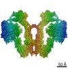
| Title | Cryo-EM of the GTP-bound human dynamin-1 polymer assembled on the membrane in the super constricted state | |||||||||
 Components Components | Dynamin-1 | |||||||||
 Keywords Keywords | ENDOCYTOSIS / HYDROLASE / dynamin family / gtpase / pleckstrin homology domain / membrane protein | |||||||||
| Function / homology |  Function and homology information Function and homology informationclathrin coat assembly involved in endocytosis / vesicle scission / synaptic vesicle budding from presynaptic endocytic zone membrane / dynamin GTPase / chromaffin granule / regulation of vesicle size / Retrograde neurotrophin signalling / Toll Like Receptor 4 (TLR4) Cascade / Formation of annular gap junctions / endosome organization ...clathrin coat assembly involved in endocytosis / vesicle scission / synaptic vesicle budding from presynaptic endocytic zone membrane / dynamin GTPase / chromaffin granule / regulation of vesicle size / Retrograde neurotrophin signalling / Toll Like Receptor 4 (TLR4) Cascade / Formation of annular gap junctions / endosome organization / Gap junction degradation / Recycling pathway of L1 / phosphatidylinositol-3,4,5-trisphosphate binding / EPH-ephrin mediated repulsion of cells / endocytic vesicle / phosphatidylinositol-4,5-bisphosphate binding / clathrin-coated pit / MHC class II antigen presentation / receptor-mediated endocytosis / protein homooligomerization / endocytosis / GDP binding / Clathrin-mediated endocytosis / presynapse / microtubule binding / protein homotetramerization / microtubule / GTPase activity / synapse / protein kinase binding / GTP binding / protein homodimerization activity / RNA binding / extracellular exosome / identical protein binding / plasma membrane / cytoplasm Similarity search - Function | |||||||||
| Biological species |  Homo sapiens (human) Homo sapiens (human) | |||||||||
| Method | ELECTRON MICROSCOPY / helical reconstruction / cryo EM / Resolution: 10.1 Å | |||||||||
 Authors Authors | Kong, L. / Wang, H. / Fang, S. / Canagarajah, B. / Kehr, A.D. / Rice, W.J. / Hinshaw, J.E. | |||||||||
| Funding support |  United States, 2items United States, 2items
| |||||||||
 Citation Citation | Journal: Nature / Year: 2018 Title: Cryo-EM of the dynamin polymer assembled on lipid membrane. Authors: Leopold Kong / Kem A Sochacki / Huaibin Wang / Shunming Fang / Bertram Canagarajah / Andrew D Kehr / William J Rice / Marie-Paule Strub / Justin W Taraska / Jenny E Hinshaw / Abstract: Membrane fission is a fundamental process in the regulation and remodelling of cell membranes. Dynamin, a large GTPase, mediates membrane fission by assembling around, constricting and cleaving the ...Membrane fission is a fundamental process in the regulation and remodelling of cell membranes. Dynamin, a large GTPase, mediates membrane fission by assembling around, constricting and cleaving the necks of budding vesicles. Here we report a 3.75 Å resolution cryo-electron microscopy structure of the membrane-associated helical polymer of human dynamin-1 in the GMPPCP-bound state. The structure defines the helical symmetry of the dynamin polymer and the positions of its oligomeric interfaces, which were validated by cell-based endocytosis assays. Compared to the lipid-free tetramer form, membrane-associated dynamin binds to the lipid bilayer with its pleckstrin homology domain (PHD) and self-assembles across the helical rungs via its guanine nucleotide-binding (GTPase) domain. Notably, interaction with the membrane and helical assembly are accommodated by a severely bent bundle signalling element (BSE), which connects the GTPase domain to the rest of the protein. The BSE conformation is asymmetric across the inter-rung GTPase interface, and is unique compared to all known nucleotide-bound states of dynamin. The structure suggests that the BSE bends as a result of forces generated from the GTPase dimer interaction that are transferred across the stalk to the PHD and lipid membrane. Mutations that disrupted the BSE kink impaired endocytosis. We also report a 10.1 Å resolution cryo-electron microscopy map of a super-constricted dynamin polymer showing localized conformational changes at the BSE and GTPase domains, induced by GTP hydrolysis, that drive membrane constriction. Together, our results provide a structural basis for the mechanism of action of dynamin on the lipid membrane. | |||||||||
| History |
|
- Structure visualization
| Movie |
Movie viewer |
|---|---|
| Structure viewer | Molecule: MolmilJmol/JSmol |
- Downloads & links
-Download
| PDBx/mmCIF format | 6dlv.cif.gz | 477.9 KB | Display | PDBx/mmCIF format |
|---|---|---|---|---|
| PDB format | pdb6dlv.ent.gz | 400 KB | Display | PDB format |
| PDBx/mmJSON format | 6dlv.json.gz | Tree view | PDBx/mmJSON format | |
| Others |  Other downloads Other downloads |
-Validation report
| Arichive directory | https://data.pdbj.org/pub/pdb/validation_reports/dl/6dlvftp://data.pdbj.org/pub/pdb/validation_reports/dl/6dlv | HTTPS FTP |
|---|
-Related structure data
| Related structure data |  7958MC  7957C  6dluC C: citing same article ( M: map data used to model this data |
|---|---|
| Similar structure data |
-Links
 PDBj
PDBj


- Assembly
| Deposited unit | 
|
|---|---|
| 1 |
|
| Symmetry | Helical symmetry: (Circular symmetry: 1 / Dyad axis: no / N subunits divisor: 1 / Num. of operations: 15 / Rise per n subunits: 14.63 Å / Rotation per n subunits: 26.14 °) |
-Components
| #1: Protein | Mass: 85859.148 Da / Num. of mol.: 4 Source method: isolated from a genetically manipulated source Source: (gene. exp.) Homo sapiens (human) / Gene: DNM1, DNM / Production host:  Trichoplusia ni (cabbage looper) / References: UniProt: Q05193, dynamin GTPase Trichoplusia ni (cabbage looper) / References: UniProt: Q05193, dynamin GTPase |
|---|
-Experimental details
-Experiment
| Experiment | Method: ELECTRON MICROSCOPY |
|---|---|
| EM experiment | Aggregation state: HELICAL ARRAY / 3D reconstruction method: helical reconstruction |
- Sample preparation
| Component | Name: GTP-bound human dynamin helical polymer encased around phosphatidylserine lipid bilayer membrane tube Type: COMPLEX / Entity ID: all / Source: RECOMBINANT |
|---|---|
| Molecular weight | Value: 98 kDa/nm / Experimental value: YES |
| Source (natural) | Organism: Homo sapiens (human) |
| Source (recombinant) | Organism: Trichoplusia ni (cabbage looper) |
| Buffer solution | pH: 7.2 |
| Specimen | Embedding applied: NO / Shadowing applied: NO / Staining applied: NO / Vitrification applied: YES |
| Vitrification | Cryogen name: ETHANE |
- Electron microscopy imaging
| Microscopy | Model: FEI TECNAI 20 |
|---|---|
| Electron gun | Electron source:  FIELD EMISSION GUN / Accelerating voltage: 200 kV / Illumination mode: FLOOD BEAM FIELD EMISSION GUN / Accelerating voltage: 200 kV / Illumination mode: FLOOD BEAM |
| Electron lens | Mode: BRIGHT FIELD |
| Image recording | Electron dose: 36 e/Å2 / Film or detector model: GATAN K2 SUMMIT (4k x 4k) |
- Processing
| CTF correction | Type: PHASE FLIPPING AND AMPLITUDE CORRECTION |
|---|---|
| Helical symmerty | Angular rotation/subunit: 26.14 ° / Axial rise/subunit: 14.63 Å / Axial symmetry: C1 |
| 3D reconstruction | Resolution: 10.1 Å / Resolution method: FSC 0.143 CUT-OFF / Num. of particles: 14322 / Symmetry type: HELICAL |
