 Movie
Movie Controller

[English] 日本語
 Yorodumi
- PDB-6d2x: Crystal structure of the GH26 domain from PbGH26-GH5A endo-beta-m... -
+ Open data

- Basic information
| Entry | Database: PDB / ID: 6d2x | ||||||
|---|---|---|---|---|---|---|---|
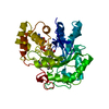
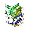
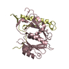
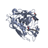
| Title | Crystal structure of the GH26 domain from PbGH26-GH5A endo-beta-mannanase/endo-beta-glucanase from Prevotella bryantii | ||||||
 Components Components | Aryl-phospho-beta-D-glucosidase BglC, GH1 family | ||||||
 Keywords Keywords | HYDROLASE / glycosyl hydrolase family 26 / glycosyl hydrolase family 5 / GH26 / GH5 / mannanase / glucanase / polysaccharide utilization locus / Prevotella bryantii | ||||||
| Function / homology |  Function and homology information Function and homology informationsubstituted mannan metabolic process / mannan endo-1,4-beta-mannosidase activity / polysaccharide catabolic process Similarity search - Function | ||||||
| Biological species |  Prevotella bryantii B14 (bacteria) Prevotella bryantii B14 (bacteria) | ||||||
| Method |  X-RAY DIFFRACTION / MOLECULAR REPLACEMENT / Resolution: 1.72 Å X-RAY DIFFRACTION / MOLECULAR REPLACEMENT / Resolution: 1.72 Å | ||||||
 Authors Authors | Stogios, P.J. / Skarina, T. / McGregor, N. / Di Leo, R. / Brumer, H. / Savchenko, A. | ||||||
| Funding support |  Canada, 1items Canada, 1items
| ||||||
 Citation Citation | Journal: To Be Published Title: Crystal structure of the GH26 domain from PbGH26-GH5A endo-beta-mannanase/endo-beta-glucanase from Prevotella bryantii Authors: McGregor, N. | ||||||
| History |
|
- Structure visualization
| Structure viewer | Molecule: MolmilJmol/JSmol |
|---|
- Downloads & links
-Download
| PDBx/mmCIF format | 6d2x.cif.gz | 167.1 KB | Display | PDBx/mmCIF format |
|---|---|---|---|---|
| PDB format | pdb6d2x.ent.gz | 127.5 KB | Display | PDB format |
| PDBx/mmJSON format | 6d2x.json.gz | Tree view | PDBx/mmJSON format | |
| Others |  Other downloads Other downloads |
-Validation report
| Arichive directory | https://data.pdbj.org/pub/pdb/validation_reports/d2/6d2xftp://data.pdbj.org/pub/pdb/validation_reports/d2/6d2x | HTTPS FTP |
|---|
-Related structure data
| Related structure data |  6d2wS S: Starting model for refinement |
|---|---|
| Similar structure data |
-Links
 PDBj
PDBj- Assembly
| Deposited unit | 
| ||||||||||||||||||
|---|---|---|---|---|---|---|---|---|---|---|---|---|---|---|---|---|---|---|---|
| 1 |
| ||||||||||||||||||
| Unit cell |
| ||||||||||||||||||
| Components on special symmetry positions |
|
-Components
| #1: Protein | Mass: 38148.188 Da / Num. of mol.: 1 Source method: isolated from a genetically manipulated source Source: (gene. exp.) Prevotella bryantii B14 (bacteria) / Gene: PBR_0368, SAMN05444375_1266 / Plasmid: pMCSG53 / Production host: | ||||
|---|---|---|---|---|---|
| #2: Chemical | ChemComp-GOL /   Mass: 92.094 Da / Num. of mol.: 1 / Source method: obtained synthetically / Formula: C3H8O3 Mass: 92.094 Da / Num. of mol.: 1 / Source method: obtained synthetically / Formula: C3H8O3 | ||||
| #3: Chemical | ChemComp-PE3 /   Mass: 634.751 Da / Num. of mol.: 5 / Source method: obtained synthetically / Formula: C28H58O15 Mass: 634.751 Da / Num. of mol.: 5 / Source method: obtained synthetically / Formula: C28H58O15#4: Chemical |   Mass: 96.063 Da / Num. of mol.: 3 / Source method: obtained synthetically / Formula: SO4 Mass: 96.063 Da / Num. of mol.: 3 / Source method: obtained synthetically / Formula: SO4#5: Water | ChemComp-HOH / |  Mass: 18.015 Da / Num. of mol.: 547 / Source method: isolated from a natural source / Formula: H2O Mass: 18.015 Da / Num. of mol.: 547 / Source method: isolated from a natural source / Formula: H2O |
-Experimental details
-Experiment
| Experiment | Method: X-RAY DIFFRACTION / Number of used crystals: 1 |
|---|
- Sample preparation
| Crystal | Density Matthews: 2.15 Å3/Da / Density % sol: 42.86 % |
|---|---|
| Crystal grow | Temperature: 298 K / Method: vapor diffusion, sitting drop / pH: 7 Details: 2 M ammonium sulfate, 2% (w/v) PEG200, 15 mg/mL protein |
-Data collection
| Diffraction | Mean temperature: 100 K |
|---|---|
| Diffraction source | Source: ROTATING ANODE / Type: RIGAKU MICROMAX-007 HF / Wavelength: 1.5418 Å |
| Detector | Type: RIGAKU RAXIS IV / Detector: IMAGE PLATE / Date: Jun 13, 2017 |
| Radiation | Protocol: SINGLE WAVELENGTH / Monochromatic (M) / Laue (L): M / Scattering type: x-ray |
| Radiation wavelength | Wavelength: 1.5418 Å / Relative weight: 1 |
| Reflection | Resolution: 1.72→30 Å / Num. obs: 35561 / % possible obs: 99.9 % / Redundancy: 6.4 % / Rmerge(I) obs: 0.082 / Rpim(I) all: 0.035 / Net I/σ(I): 36.3 |
| Reflection shell | Resolution: 1.72→1.75 Å / Redundancy: 6.3 % / Rmerge(I) obs: 1.142 / Mean I/σ(I) obs: 2.14 / Num. unique all: 1761 / CC1/2: 0.607 / Rpim(I) all: 0.492 / % possible all: 100 |
- Processing
| Software |
| ||||||||||||||||||||||||||||||||||||||||||||||||||||||||||||||||||||||||||||||||||||||||||||||||||||||||||||||||||||||||||||||||||||||||||||||||||||||
|---|---|---|---|---|---|---|---|---|---|---|---|---|---|---|---|---|---|---|---|---|---|---|---|---|---|---|---|---|---|---|---|---|---|---|---|---|---|---|---|---|---|---|---|---|---|---|---|---|---|---|---|---|---|---|---|---|---|---|---|---|---|---|---|---|---|---|---|---|---|---|---|---|---|---|---|---|---|---|---|---|---|---|---|---|---|---|---|---|---|---|---|---|---|---|---|---|---|---|---|---|---|---|---|---|---|---|---|---|---|---|---|---|---|---|---|---|---|---|---|---|---|---|---|---|---|---|---|---|---|---|---|---|---|---|---|---|---|---|---|---|---|---|---|---|---|---|---|---|---|---|---|
| Refinement | Method to determine structure: MOLECULAR REPLACEMENT Starting model: 6D2W Resolution: 1.72→27.243 Å / SU ML: 0.18 / Cross valid method: FREE R-VALUE / σ(F): 1.34 / Phase error: 19.65 / Stereochemistry target values: ML
| ||||||||||||||||||||||||||||||||||||||||||||||||||||||||||||||||||||||||||||||||||||||||||||||||||||||||||||||||||||||||||||||||||||||||||||||||||||||
| Solvent computation | Shrinkage radii: 0.9 Å / VDW probe radii: 1.11 Å / Solvent model: FLAT BULK SOLVENT MODEL | ||||||||||||||||||||||||||||||||||||||||||||||||||||||||||||||||||||||||||||||||||||||||||||||||||||||||||||||||||||||||||||||||||||||||||||||||||||||
| Refinement step | Cycle: LAST / Resolution: 1.72→27.243 Å
| ||||||||||||||||||||||||||||||||||||||||||||||||||||||||||||||||||||||||||||||||||||||||||||||||||||||||||||||||||||||||||||||||||||||||||||||||||||||
| Refine LS restraints |
| ||||||||||||||||||||||||||||||||||||||||||||||||||||||||||||||||||||||||||||||||||||||||||||||||||||||||||||||||||||||||||||||||||||||||||||||||||||||
| LS refinement shell |
| ||||||||||||||||||||||||||||||||||||||||||||||||||||||||||||||||||||||||||||||||||||||||||||||||||||||||||||||||||||||||||||||||||||||||||||||||||||||
| Refinement TLS params. | Method: refined / Refine-ID: X-RAY DIFFRACTION
| ||||||||||||||||||||||||||||||||||||||||||||||||||||||||||||||||||||||||||||||||||||||||||||||||||||||||||||||||||||||||||||||||||||||||||||||||||||||
| Refinement TLS group |
|
