 Movie
Movie Controller

[English] 日本語
 Yorodumi
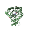
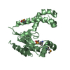
- PDB-6cuj: Crystal structure of the C-terminal domain of neisserial heparin ... -
+ Open data

- Basic information
| Entry | Database: PDB / ID: 6cuj | ||||||
|---|---|---|---|---|---|---|---|
| Title | Crystal structure of the C-terminal domain of neisserial heparin binding antigen (NHBA) | ||||||
 Components Components | Gna2132 | ||||||
 Keywords Keywords | LIPID BINDING PROTEIN / Vaccine / Neisseria / Meningitis / Disorder | ||||||
| Function / homology | Transferrin-binding protein B, C-lobe/N-lobe beta barrel domain / C-lobe and N-lobe beta barrels of Tf-binding protein B / Outer membrane protein/outer membrane enzyme PagP, beta-barrel / cell outer membrane / heparin binding / Prokaryotic membrane lipoprotein lipid attachment site profile. / cell adhesion / DNA binding / Neisserial heparin binding antigen Function and homology information Function and homology information | ||||||
| Biological species |  Neisseria meningitidis (bacteria) Neisseria meningitidis (bacteria) | ||||||
| Method |  X-RAY DIFFRACTION / SYNCHROTRON / MOLECULAR REPLACEMENT / Resolution: 1.805 Å X-RAY DIFFRACTION / SYNCHROTRON / MOLECULAR REPLACEMENT / Resolution: 1.805 Å | ||||||
 Authors Authors | Malito, E. / Spraggon, G. | ||||||
 Citation Citation | Journal: PLoS ONE / Year: 2018 Title: Structures of NHBA elucidate a broadly conserved epitope identified by a vaccine induced antibody. Authors: Maritan, M. / Veggi, D. / Cozzi, R. / Dello Iacono, L. / Bartolini, E. / Lo Surdo, P. / Maruggi, G. / Spraggon, G. / Bottomley, M.J. / Malito, E. | ||||||
| History |
|
- Structure visualization
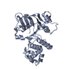
| Structure viewer | Molecule: MolmilJmol/JSmol |
|---|
- Downloads & links
-Download
| PDBx/mmCIF format | 6cuj.cif.gz | 71.8 KB | Display | PDBx/mmCIF format |
|---|---|---|---|---|
| PDB format | pdb6cuj.ent.gz | 52.8 KB | Display | PDB format |
| PDBx/mmJSON format | 6cuj.json.gz | Tree view | PDBx/mmJSON format | |
| Others |  Other downloads Other downloads |
-Validation report
| Arichive directory | https://data.pdbj.org/pub/pdb/validation_reports/cu/6cujftp://data.pdbj.org/pub/pdb/validation_reports/cu/6cuj | HTTPS FTP |
|---|
-Related structure data
| Related structure data | 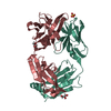 5nyxC 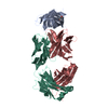 5o1rC  2lfuS S: Starting model for refinement C: citing same article ( |
|---|---|
| Similar structure data |


-Links
 PDBj
PDBj


- Assembly
| Deposited unit | 
| ||||||||
|---|---|---|---|---|---|---|---|---|---|
| 1 |
| ||||||||
| Unit cell |
|
-Components
| #1: Protein | Mass: 17377.043 Da / Num. of mol.: 2 / Fragment: UNP Residues 133-427 Source method: isolated from a genetically manipulated source Source: (gene. exp.) Neisseria meningitidis (bacteria) / Gene: gna2132, nhba, A6J54_02980 / Production host: #2: Water | ChemComp-HOH / |  Mass: 18.015 Da / Num. of mol.: 121 / Source method: isolated from a natural source / Formula: H2O Mass: 18.015 Da / Num. of mol.: 121 / Source method: isolated from a natural source / Formula: H2OSequence details | The full sequence MAANGGSNFGRVDLANGVLIDGPSQNITLTHSKGDSSNGDNLLDEEAPSKSEFENLNESERIEKYKKDGKSDKFTNLVAT ...The full sequence MAANGGSNFG | |
|---|
-Experimental details
-Experiment
| Experiment | Method: X-RAY DIFFRACTION / Number of used crystals: 1 |
|---|
- Sample preparation
| Crystal | Density Matthews: 2.49 Å3/Da / Density % sol: 50 % |
|---|---|
| Crystal grow | Temperature: 293 K / Method: vapor diffusion, sitting drop / Details: 20% PEG 3,350, 0.2 M Sodium thiocyanate |
-Data collection
| Diffraction | Mean temperature: 80 K |
|---|---|
| Diffraction source | Source: SYNCHROTRON / Site: ESRF  / Beamline: ID29 / Wavelength: 0.96862 Å / Beamline: ID29 / Wavelength: 0.96862 Å |
| Detector | Type: DECTRIS PILATUS 6M-F / Detector: PIXEL / Date: Aug 29, 2014 |
| Radiation | Protocol: SINGLE WAVELENGTH / Monochromatic (M) / Laue (L): M / Scattering type: x-ray |
| Radiation wavelength | Wavelength: 0.96862 Å / Relative weight: 1 |
| Reflection | Resolution: 1.8→46 Å / Num. obs: 31127 / % possible obs: 99.9 % / Redundancy: 4.6 % / Biso Wilson estimate: 32.8 Å2 / CC1/2: 0.99 / Rrim(I) all: 0.06 / Rsym value: 0.06 / Net I/σ(I): 13 |
| Reflection shell | Resolution: 1.8→1.91 Å / Redundancy: 4.7 % / Mean I/σ(I) obs: 2 / Num. unique obs: 5025 / CC1/2: 0.84 / Rsym value: 0.47 / % possible all: 99.9 |
- Processing
| Software |
| ||||||||||||||||||||||||||||||||||||||||||||||||||||||||||||||||||||||||||||||||||||
|---|---|---|---|---|---|---|---|---|---|---|---|---|---|---|---|---|---|---|---|---|---|---|---|---|---|---|---|---|---|---|---|---|---|---|---|---|---|---|---|---|---|---|---|---|---|---|---|---|---|---|---|---|---|---|---|---|---|---|---|---|---|---|---|---|---|---|---|---|---|---|---|---|---|---|---|---|---|---|---|---|---|---|---|---|---|
| Refinement | Method to determine structure: MOLECULAR REPLACEMENT Starting model: 2LFU Resolution: 1.805→46 Å / SU ML: 0.22 / Cross valid method: FREE R-VALUE / σ(F): 1.38 / Phase error: 23.18
| ||||||||||||||||||||||||||||||||||||||||||||||||||||||||||||||||||||||||||||||||||||
| Solvent computation | Shrinkage radii: 0.9 Å / VDW probe radii: 1.11 Å | ||||||||||||||||||||||||||||||||||||||||||||||||||||||||||||||||||||||||||||||||||||
| Refinement step | Cycle: LAST / Resolution: 1.805→46 Å
| ||||||||||||||||||||||||||||||||||||||||||||||||||||||||||||||||||||||||||||||||||||
| Refine LS restraints |
| ||||||||||||||||||||||||||||||||||||||||||||||||||||||||||||||||||||||||||||||||||||
| LS refinement shell |
|
