 Movie
Movie Controller


+ Open data

- Basic information
| Entry | Database: PDB / ID: 6cfy | ||||||
|---|---|---|---|---|---|---|---|
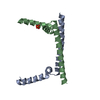
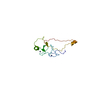
| Title | Bosea sp Root 381 apo GapR structure | ||||||
 Components Components | UPF0335 protein ASE63_04290 | ||||||
 Keywords Keywords | DNA BINDING PROTEIN / Caulobacter / NAP / DNA binding / topoisomerase | ||||||
| Function / homology | GapR-like / GapR-like, DNA-binding domain / GapR-like, DNA-binding domain / DNA binding / UPF0335 protein ASE63_04290 Function and homology information Function and homology information | ||||||
| Biological species |  Bosea sp. Root381 (bacteria) Bosea sp. Root381 (bacteria) | ||||||
| Method |  X-RAY DIFFRACTION / SYNCHROTRON / SAD / Resolution: 2.4 Å X-RAY DIFFRACTION / SYNCHROTRON / SAD / Resolution: 2.4 Å | ||||||
 Authors Authors | Schumacherr, M.A. | ||||||
 Citation Citation | Journal: Cell / Year: 2018 Title: A Bacterial Chromosome Structuring Protein Binds Overtwisted DNA to Stimulate Type II Topoisomerases and Enable DNA Replication. Authors: Guo, M.S. / Haakonsen, D.L. / Zeng, W. / Schumacher, M.A. / Laub, M.T. | ||||||
| History |
|
- Structure visualization
| Structure viewer | Molecule: MolmilJmol/JSmol |
|---|
- Downloads & links
-Download
| PDBx/mmCIF format | 6cfy.cif.gz | 41.4 KB | Display | PDBx/mmCIF format |
|---|---|---|---|---|
| PDB format | pdb6cfy.ent.gz | 28.4 KB | Display | PDB format |
| PDBx/mmJSON format | 6cfy.json.gz | Tree view | PDBx/mmJSON format | |
| Others |  Other downloads Other downloads |
-Validation report
| Arichive directory | https://data.pdbj.org/pub/pdb/validation_reports/cf/6cfyftp://data.pdbj.org/pub/pdb/validation_reports/cf/6cfy | HTTPS FTP |
|---|
-Related structure data


-Links
 PDBj
PDBj
- Assembly
| Deposited unit | 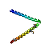
| ||||||||
|---|---|---|---|---|---|---|---|---|---|
| 1 | 
| ||||||||
| Unit cell |
|
-Components
| #1: Protein | Mass: 9287.882 Da / Num. of mol.: 1 / Mutation: L48M, I54M, L73M Source method: isolated from a genetically manipulated source Source: (gene. exp.) Bosea sp. Root381 (bacteria) / Gene: ASE63_04290 / Production host: |
|---|---|
| #2: Water | ChemComp-HOH /  Mass: 18.015 Da / Num. of mol.: 5 / Source method: isolated from a natural source / Formula: H2O Mass: 18.015 Da / Num. of mol.: 5 / Source method: isolated from a natural source / Formula: H2O |
| Has protein modification | Y |
-Experimental details
-Experiment
| Experiment | Method: X-RAY DIFFRACTION / Number of used crystals: 1 |
|---|
- Sample preparation
| Crystal | Density Matthews: 4.85 Å3/Da / Density % sol: 74.65 % |
|---|---|
| Crystal grow | Temperature: 298 K / Method: vapor diffusion, hanging drop / Details: 15% MPD, 10 mM MgCl2, HEPES (7.5) 0.001 M spermine |
-Data collection
| Diffraction | Mean temperature: 273 K |
|---|---|
| Diffraction source | Source: SYNCHROTRON / Site: ALS  / Beamline: 8.3.1 / Wavelength: 0.979 Å / Beamline: 8.3.1 / Wavelength: 0.979 Å |
| Detector | Type: DECTRIS PILATUS3 S 6M / Detector: PIXEL / Date: Dec 16, 2017 |
| Radiation | Protocol: SINGLE WAVELENGTH / Monochromatic (M) / Laue (L): M / Scattering type: x-ray |
| Radiation wavelength | Wavelength: 0.979 Å / Relative weight: 1 |
| Reflection | Resolution: 2.4→38.87 Å / Num. obs: 12187 / % possible obs: 100 % / Redundancy: 19.1 % / CC1/2: 0.997 / Rpim(I) all: 0.041 / Rrim(I) all: 0.122 / Rsym value: 0.099 / Net I/σ(I): 11.2 |
| Reflection shell | Resolution: 2.4→2.53 Å / Num. unique obs: 1213 / CC1/2: 0.319 / % possible all: 99.9 |
-Phasing
| Phasing | Method: SAD |
|---|
- Processing
| Software |
| ||||||||||||||||||||||||||||||||||||||||||||||||||||||||||||
|---|---|---|---|---|---|---|---|---|---|---|---|---|---|---|---|---|---|---|---|---|---|---|---|---|---|---|---|---|---|---|---|---|---|---|---|---|---|---|---|---|---|---|---|---|---|---|---|---|---|---|---|---|---|---|---|---|---|---|---|---|---|
| Refinement | Method to determine structure: SAD / Resolution: 2.4→38.8 Å / SU ML: 0.44 / Cross valid method: THROUGHOUT / σ(F): 0.79 / Phase error: 22.03
| ||||||||||||||||||||||||||||||||||||||||||||||||||||||||||||
| Solvent computation | Shrinkage radii: 0.95 Å / VDW probe radii: 1.2 Å / Bsol: 81.292 Å2 / ksol: 0.4 e/Å3 | ||||||||||||||||||||||||||||||||||||||||||||||||||||||||||||
| Displacement parameters | Biso max: 140.06 Å2 / Biso mean: 69.35 Å2 / Biso min: 41.4 Å2
| ||||||||||||||||||||||||||||||||||||||||||||||||||||||||||||
| Refinement step | Cycle: final / Resolution: 2.4→38.8 Å
| ||||||||||||||||||||||||||||||||||||||||||||||||||||||||||||
| Refine LS restraints |
| ||||||||||||||||||||||||||||||||||||||||||||||||||||||||||||
| LS refinement shell | Refine-ID: X-RAY DIFFRACTION / Rfactor Rfree error: 0 / Total num. of bins used: 9 / % reflection obs: 100 %
| ||||||||||||||||||||||||||||||||||||||||||||||||||||||||||||
| Refinement TLS params. | Method: refined / Origin x: -0.8545 Å / Origin y: -16.4547 Å / Origin z: 9.7667 Å
| ||||||||||||||||||||||||||||||||||||||||||||||||||||||||||||
| Refinement TLS group |
|
