 Movie
Movie Controller


+ Open data

- Basic information
| Entry | Database: PDB / ID: 6bqu | |||||||||
|---|---|---|---|---|---|---|---|---|---|---|
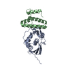
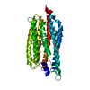
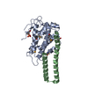
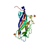
| Title | Human GR (418-507) in complex with monomeric DNA binding site | |||||||||
 Components Components |
| |||||||||
 Keywords Keywords | DNA BINDING PROTEIN/DNA / Glucocorticoid receptor Monomeric binding site / DNA BINDING PROTEIN / DNA BINDING PROTEIN-DNA complex | |||||||||
| Function / homology |  Function and homology information Function and homology informationRegulation of NPAS4 gene transcription / regulation of glucocorticoid biosynthetic process / nuclear glucocorticoid receptor activity / steroid hormone binding / PTK6 Expression / glucocorticoid metabolic process / neuroinflammatory response / response to cortisol / mammary gland duct morphogenesis / microglia differentiation ...Regulation of NPAS4 gene transcription / regulation of glucocorticoid biosynthetic process / nuclear glucocorticoid receptor activity / steroid hormone binding / PTK6 Expression / glucocorticoid metabolic process / neuroinflammatory response / response to cortisol / mammary gland duct morphogenesis / microglia differentiation / astrocyte differentiation / maternal behavior / regulation of gluconeogenesis / adrenal gland development / cellular response to glucocorticoid stimulus / cellular response to steroid hormone stimulus / motor behavior / nuclear receptor-mediated steroid hormone signaling pathway / estrogen response element binding / cellular response to dexamethasone stimulus / FOXO-mediated transcription of oxidative stress, metabolic and neuronal genes / cellular response to transforming growth factor beta stimulus / core promoter sequence-specific DNA binding / steroid binding / HSP90 chaperone cycle for steroid hormone receptors (SHR) in the presence of ligand / TBP-class protein binding / RNA polymerase II transcription regulatory region sequence-specific DNA binding / SUMOylation of intracellular receptors / promoter-specific chromatin binding / chromosome segregation / synaptic transmission, glutamatergic / Hsp90 protein binding / Nuclear Receptor transcription pathway / positive regulation of miRNA transcription / response to wounding / DNA-binding transcription repressor activity, RNA polymerase II-specific / nuclear receptor activity / spindle / sequence-specific double-stranded DNA binding / Regulation of RUNX2 expression and activity / positive regulation of neuron apoptotic process / chromatin organization / DNA-binding transcription activator activity, RNA polymerase II-specific / Potential therapeutics for SARS / gene expression / DNA-binding transcription factor activity, RNA polymerase II-specific / nuclear speck / RNA polymerase II cis-regulatory region sequence-specific DNA binding / DNA-binding transcription factor activity / mitochondrial matrix / cell division / negative regulation of DNA-templated transcription / apoptotic process / regulation of transcription by RNA polymerase II / regulation of DNA-templated transcription / centrosome / synapse / protein kinase binding / chromatin / negative regulation of transcription by RNA polymerase II / signal transduction / positive regulation of transcription by RNA polymerase II / protein-containing complex / RNA binding / zinc ion binding / nucleoplasm / membrane / identical protein binding / nucleus / cytoplasm / cytosol Similarity search - Function | |||||||||
| Biological species |  Homo sapiens (human) Homo sapiens (human)synthetic construct (others) | |||||||||
| Method |  X-RAY DIFFRACTION / SYNCHROTRON / MOLECULAR REPLACEMENT / Resolution: 2.5 Å X-RAY DIFFRACTION / SYNCHROTRON / MOLECULAR REPLACEMENT / Resolution: 2.5 Å | |||||||||
 Authors Authors | Pufall, M.A. | |||||||||
| Funding support |  United States, 2items United States, 2items
| |||||||||
 Citation Citation | Journal: To Be Published Title: General and sequence-specific roles for DNA in glucocorticoid receptor DNA-binding stoichiometry Authors: Pufall, M.A. | |||||||||
| History |
|
- Structure visualization
| Structure viewer | Molecule: MolmilJmol/JSmol |
|---|
- Downloads & links
-Download
| PDBx/mmCIF format | 6bqu.cif.gz | 140.3 KB | Display | PDBx/mmCIF format |
|---|---|---|---|---|
| PDB format | pdb6bqu.ent.gz | 109.7 KB | Display | PDB format |
| PDBx/mmJSON format | 6bqu.json.gz | Tree view | PDBx/mmJSON format | |
| Others |  Other downloads Other downloads |
-Validation report
| Arichive directory | https://data.pdbj.org/pub/pdb/validation_reports/bq/6bquftp://data.pdbj.org/pub/pdb/validation_reports/bq/6bqu | HTTPS FTP |
|---|
-Related structure data
| Related structure data |  6bseC  6bsfC  3g99S S: Starting model for refinement C: citing same article ( |
|---|---|
| Similar structure data |


-Links
 PDBj
PDBj


- Assembly
| Deposited unit | 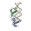
| ||||||||
|---|---|---|---|---|---|---|---|---|---|
| 1 |
| ||||||||
| Unit cell |
|
-Components
| #1: Protein | Mass: 8074.493 Da / Num. of mol.: 2 Source method: isolated from a genetically manipulated source Source: (gene. exp.) Homo sapiens (human) / Gene: NR3C1, GRL / Production host:  #2: DNA chain | | Mass: 4897.203 Da / Num. of mol.: 1 / Source method: obtained synthetically / Source: (synth.) synthetic construct (others) #3: DNA chain | | Mass: 4897.203 Da / Num. of mol.: 1 / Source method: obtained synthetically / Source: (synth.) synthetic construct (others) #4: Chemical | ChemComp-ZN /   Mass: 65.409 Da / Num. of mol.: 4 / Source method: obtained synthetically / Formula: Zn Mass: 65.409 Da / Num. of mol.: 4 / Source method: obtained synthetically / Formula: Zn#5: Water | ChemComp-HOH / |  Mass: 18.015 Da / Num. of mol.: 10 / Source method: isolated from a natural source / Formula: H2O Mass: 18.015 Da / Num. of mol.: 10 / Source method: isolated from a natural source / Formula: H2O |
|---|
-Experimental details
-Experiment
| Experiment | Method: X-RAY DIFFRACTION / Number of used crystals: 1 |
|---|
- Sample preparation
| Crystal | Density Matthews: 3.39 Å3/Da / Density % sol: 63.68 % |
|---|---|
| Crystal grow | Temperature: 293 K / Method: vapor diffusion, hanging drop Details: 0.005 M Magnesium sulfate heptahydrate, 0.05 M MES monohydrate, pH 6.0, 5 percent w/v, Polyethylene glycol 4,000 |
-Data collection
| Diffraction | Mean temperature: 100 K |
|---|---|
| Diffraction source | Source: SYNCHROTRON / Site: ALS / Beamline: 4.2.2 / Wavelength: 1 Å |
| Detector | Type: NOIR-1 / Detector: CCD / Date: Dec 19, 2016 |
| Radiation | Protocol: SINGLE WAVELENGTH / Monochromatic (M) / Laue (L): M / Scattering type: x-ray |
| Radiation wavelength | Wavelength: 1 Å / Relative weight: 1 |
| Reflection | Resolution: 2.5→51.952 Å / Num. obs: 12750 / % possible obs: 99.66 % / Redundancy: 6.8 % / Biso Wilson estimate: 40.405 Å2 / CC1/2: 0.999 / Rmerge(I) obs: 0.06019 / Net I/σ(I): 16.2 |
| Reflection shell | Resolution: 2.5→2.75 Å / Redundancy: 6.9 % / Rmerge(I) obs: 0.8355 / Num. unique all: 1246 / Num. unique obs: 3122 / CC1/2: 0.821 / % possible all: 100 |
- Processing
| Software |
| |||||||||||||||||||||||||||||||||||||||||||||||||||||||||||||||||||||||||||||||||||||||||||||||||||||||||||||||||||||||||||||||||||||||||||||||||||||||||||||||||||||||||||||||||||||||||||||||||||||||||||||||||||||||||||||||||||||||||||||||||||||||||||||||||||||||||||||||||||||||||||||||||||||||||||||||||||||||||||||||||||||
|---|---|---|---|---|---|---|---|---|---|---|---|---|---|---|---|---|---|---|---|---|---|---|---|---|---|---|---|---|---|---|---|---|---|---|---|---|---|---|---|---|---|---|---|---|---|---|---|---|---|---|---|---|---|---|---|---|---|---|---|---|---|---|---|---|---|---|---|---|---|---|---|---|---|---|---|---|---|---|---|---|---|---|---|---|---|---|---|---|---|---|---|---|---|---|---|---|---|---|---|---|---|---|---|---|---|---|---|---|---|---|---|---|---|---|---|---|---|---|---|---|---|---|---|---|---|---|---|---|---|---|---|---|---|---|---|---|---|---|---|---|---|---|---|---|---|---|---|---|---|---|---|---|---|---|---|---|---|---|---|---|---|---|---|---|---|---|---|---|---|---|---|---|---|---|---|---|---|---|---|---|---|---|---|---|---|---|---|---|---|---|---|---|---|---|---|---|---|---|---|---|---|---|---|---|---|---|---|---|---|---|---|---|---|---|---|---|---|---|---|---|---|---|---|---|---|---|---|---|---|---|---|---|---|---|---|---|---|---|---|---|---|---|---|---|---|---|---|---|---|---|---|---|---|---|---|---|---|---|---|---|---|---|---|---|---|---|---|---|---|---|---|---|---|---|---|---|---|---|---|---|---|---|---|---|---|---|---|---|---|---|---|---|---|---|---|---|---|---|---|---|---|---|---|---|---|---|---|---|---|---|---|---|---|---|---|---|---|---|---|---|---|---|---|---|---|---|
| Refinement | Method to determine structure: MOLECULAR REPLACEMENT Starting model: 3G99 Resolution: 2.5→51.952 Å / SU ML: 0.35 / Cross valid method: FREE R-VALUE / σ(F): 1.34 / Phase error: 28.2
| |||||||||||||||||||||||||||||||||||||||||||||||||||||||||||||||||||||||||||||||||||||||||||||||||||||||||||||||||||||||||||||||||||||||||||||||||||||||||||||||||||||||||||||||||||||||||||||||||||||||||||||||||||||||||||||||||||||||||||||||||||||||||||||||||||||||||||||||||||||||||||||||||||||||||||||||||||||||||||||||||||||
| Solvent computation | Shrinkage radii: 0.9 Å / VDW probe radii: 1.11 Å | |||||||||||||||||||||||||||||||||||||||||||||||||||||||||||||||||||||||||||||||||||||||||||||||||||||||||||||||||||||||||||||||||||||||||||||||||||||||||||||||||||||||||||||||||||||||||||||||||||||||||||||||||||||||||||||||||||||||||||||||||||||||||||||||||||||||||||||||||||||||||||||||||||||||||||||||||||||||||||||||||||||
| Refinement step | Cycle: LAST / Resolution: 2.5→51.952 Å
| |||||||||||||||||||||||||||||||||||||||||||||||||||||||||||||||||||||||||||||||||||||||||||||||||||||||||||||||||||||||||||||||||||||||||||||||||||||||||||||||||||||||||||||||||||||||||||||||||||||||||||||||||||||||||||||||||||||||||||||||||||||||||||||||||||||||||||||||||||||||||||||||||||||||||||||||||||||||||||||||||||||
| Refine LS restraints |
| |||||||||||||||||||||||||||||||||||||||||||||||||||||||||||||||||||||||||||||||||||||||||||||||||||||||||||||||||||||||||||||||||||||||||||||||||||||||||||||||||||||||||||||||||||||||||||||||||||||||||||||||||||||||||||||||||||||||||||||||||||||||||||||||||||||||||||||||||||||||||||||||||||||||||||||||||||||||||||||||||||||
| LS refinement shell |
| |||||||||||||||||||||||||||||||||||||||||||||||||||||||||||||||||||||||||||||||||||||||||||||||||||||||||||||||||||||||||||||||||||||||||||||||||||||||||||||||||||||||||||||||||||||||||||||||||||||||||||||||||||||||||||||||||||||||||||||||||||||||||||||||||||||||||||||||||||||||||||||||||||||||||||||||||||||||||||||||||||||
| Refinement TLS params. | Method: refined / Refine-ID: X-RAY DIFFRACTION
| |||||||||||||||||||||||||||||||||||||||||||||||||||||||||||||||||||||||||||||||||||||||||||||||||||||||||||||||||||||||||||||||||||||||||||||||||||||||||||||||||||||||||||||||||||||||||||||||||||||||||||||||||||||||||||||||||||||||||||||||||||||||||||||||||||||||||||||||||||||||||||||||||||||||||||||||||||||||||||||||||||||
| Refinement TLS group |
|
