 Movie
Movie Controller


+ Open data

- Basic information
| Entry | Database: PDB / ID: 6azd | ||||||
|---|---|---|---|---|---|---|---|
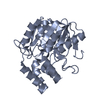
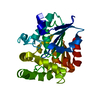
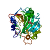
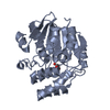
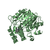
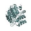
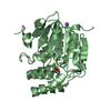
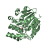
| Title | Crystal structure of Physcomitrella patens KAI2-like H | ||||||
 Components Components | PpKAI2-like H | ||||||
 Keywords Keywords | HYDROLASE / alpha/beta hydrolase / strigolactone binding | ||||||
| Function / homology | alpha/beta hydrolase fold / Alpha/beta hydrolase fold-1 / Alpha/Beta hydrolase fold, catalytic domain / Alpha/Beta hydrolase fold / Rossmann fold / 3-Layer(aba) Sandwich / Alpha Beta / AB hydrolase-1 domain-containing protein Function and homology information Function and homology information | ||||||
| Biological species |  Physcomitrella patens subsp. patens (plant) Physcomitrella patens subsp. patens (plant) | ||||||
| Method |  X-RAY DIFFRACTION / SYNCHROTRON / MOLECULAR REPLACEMENT / molecular replacement / Resolution: 1.97010740396 Å X-RAY DIFFRACTION / SYNCHROTRON / MOLECULAR REPLACEMENT / molecular replacement / Resolution: 1.97010740396 Å | ||||||
 Authors Authors | Burger, M. / Lee, H.J. / Chory, J. | ||||||
 Citation Citation | Journal: Cell Rep / Year: 2019 Title: Structural Basis of Karrikin and Non-natural Strigolactone Perception in Physcomitrella patens. Authors: Burger, M. / Mashiguchi, K. / Lee, H.J. / Nakano, M. / Takemoto, K. / Seto, Y. / Yamaguchi, S. / Chory, J. | ||||||
| History |
|
- Structure visualization
| Structure viewer | Molecule: MolmilJmol/JSmol |
|---|
- Downloads & links
-Download
| PDBx/mmCIF format | 6azd.cif.gz | 81.9 KB | Display | PDBx/mmCIF format |
|---|---|---|---|---|
| PDB format | pdb6azd.ent.gz | 48.4 KB | Display | PDB format |
| PDBx/mmJSON format | 6azd.json.gz | Tree view | PDBx/mmJSON format | |
| Others |  Other downloads Other downloads |
-Validation report
| Summary document | 6azd_validation.pdf.gz | 419.4 KB | Display | wwPDB validaton report |
|---|---|---|---|---|
| Full document | 6azd_full_validation.pdf.gz | 421.8 KB | Display | |
| Data in XML | 6azd_validation.xml.gz | 12.8 KB | Display | |
| Data in CIF | 6azd_validation.cif.gz | 18 KB | Display | |
| Arichive directory | https://data.pdbj.org/pub/pdb/validation_reports/az/6azdftp://data.pdbj.org/pub/pdb/validation_reports/az/6azd | HTTPS FTP |
-Related structure data
| Related structure data | 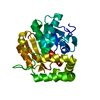 6atxC  6azbC  6azcC 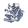 4jypS S: Starting model for refinement C: citing same article ( |
|---|---|
| Similar structure data |


-Links
 PDBj
PDBj


- Assembly
| Deposited unit | 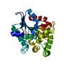
| ||||||||||||
|---|---|---|---|---|---|---|---|---|---|---|---|---|---|
| 1 |
| ||||||||||||
| Unit cell |
|
-Components
| #1: Protein | Mass: 29864.607 Da / Num. of mol.: 1 Source method: isolated from a genetically manipulated source Source: (gene. exp.) Physcomitrella patens subsp. patens (plant)Gene: PHYPADRAFT_184560 / Plasmid: pGEX4T3 / Production host:  |
|---|---|
| #2: Water | ChemComp-HOH /  Mass: 18.015 Da / Num. of mol.: 155 / Source method: isolated from a natural source / Formula: H2O Mass: 18.015 Da / Num. of mol.: 155 / Source method: isolated from a natural source / Formula: H2O |
-Experimental details
-Experiment
| Experiment | Method: X-RAY DIFFRACTION / Number of used crystals: 1 |
|---|
- Sample preparation
| Crystal | Density Matthews: 2.27 Å3/Da / Density % sol: 45.75 % |
|---|---|
| Crystal grow | Temperature: 298 K / Method: vapor diffusion, hanging drop / pH: 6.5 / Details: 1.8 M ammonium sulfate, 0.1 M MES 6.5, 5% PEG400 |
-Data collection
| Diffraction | Mean temperature: 100 K |
|---|---|
| Diffraction source | Source: SYNCHROTRON / Site: ALS  / Beamline: 8.2.1 / Wavelength: 1 Å / Beamline: 8.2.1 / Wavelength: 1 Å |
| Detector | Type: ADSC QUANTUM 315r / Detector: CCD / Date: Aug 8, 2016 / Details: mirrors |
| Radiation | Monochromator: double crystal Si(111) / Protocol: SINGLE WAVELENGTH / Monochromatic (M) / Laue (L): M / Scattering type: x-ray |
| Radiation wavelength | Wavelength: 1 Å / Relative weight: 1 |
| Reflection | Resolution: 1.97→37.1 Å / Num. obs: 18745 / % possible obs: 98.31 % / Redundancy: 3.5 % / CC1/2: 0.982 / Rmerge(I) obs: 0.1674 / Rpim(I) all: 0.1065 / Net I/σ(I): 8.17 |
| Reflection shell | Resolution: 1.97→2.04 Å / Redundancy: 3.4 % / Rmerge(I) obs: 0.4449 / Mean I/σ(I) obs: 3.85 / Num. unique obs: 1833 / CC1/2: 0.549 / Rpim(I) all: 0.285 / % possible all: 97.66 |
-Phasing
| Phasing | Method: molecular replacement |
|---|
- Processing
| Software |
| ||||||||||||||||||||||||||||||||||||||||||||||||||||||||
|---|---|---|---|---|---|---|---|---|---|---|---|---|---|---|---|---|---|---|---|---|---|---|---|---|---|---|---|---|---|---|---|---|---|---|---|---|---|---|---|---|---|---|---|---|---|---|---|---|---|---|---|---|---|---|---|---|---|
| Refinement | Method to determine structure: MOLECULAR REPLACEMENT Starting model: PDB entry 4JYP Resolution: 1.97010740396→37.0182176364 Å / SU ML: 0.277465883878 / Cross valid method: FREE R-VALUE / σ(F): 1.3487061327 / Phase error: 24.1330848868
| ||||||||||||||||||||||||||||||||||||||||||||||||||||||||
| Solvent computation | Shrinkage radii: 0.9 Å / VDW probe radii: 1.11 Å | ||||||||||||||||||||||||||||||||||||||||||||||||||||||||
| Displacement parameters | Biso mean: 14.9028463926 Å2 | ||||||||||||||||||||||||||||||||||||||||||||||||||||||||
| Refinement step | Cycle: LAST / Resolution: 1.97010740396→37.0182176364 Å
| ||||||||||||||||||||||||||||||||||||||||||||||||||||||||
| Refine LS restraints |
| ||||||||||||||||||||||||||||||||||||||||||||||||||||||||
| LS refinement shell |
|
