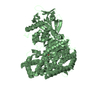
 Movie
Movie Controller

[English] 日本語
 Yorodumi
- PDB-6apz: Crystal structure of Mycobacterium tuberculosis malate synthase i... -
+ Open data

- Basic information
| Entry | Database: PDB / ID: 6apz | ||||||
|---|---|---|---|---|---|---|---|
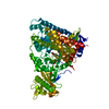
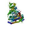
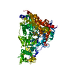
| Title | Crystal structure of Mycobacterium tuberculosis malate synthase in complex with 3-hydroxy-phenyldiketoacid | ||||||
 Components Components | Malate synthase G | ||||||
 Keywords Keywords | TRANSFERASE / Acetyltransferase / Structural Genomics / TB Structural Genomics Consortium / TBSGC | ||||||
| Function / homology |  Function and homology information Function and homology informationmalate synthase / malate synthase activity / glyoxylate catabolic process / glyoxylate cycle / tricarboxylic acid cycle / magnesium ion binding / cell surface / cytosol Similarity search - Function | ||||||
| Biological species |   Mycobacterium tuberculosis (bacteria) Mycobacterium tuberculosis (bacteria) | ||||||
| Method |  X-RAY DIFFRACTION / MOLECULAR REPLACEMENT / Resolution: 2.254 Å X-RAY DIFFRACTION / MOLECULAR REPLACEMENT / Resolution: 2.254 Å | ||||||
 Authors Authors | Krieger, I.V. / Sacchettini, J.C. / TB Structural Genomics Consortium (TBSGC) | ||||||
| Funding support |  United States, 1items United States, 1items
| ||||||
 Citation Citation | Journal: J Chem Inf Model / Year: 2018 Title: Anion-pi Interactions in Computer-Aided Drug Design: Modeling the Inhibition of Malate Synthase by Phenyl-Diketo Acids. Authors: Ellenbarger, J.F. / Krieger, I.V. / Huang, H.L. / Gomez-Coca, S. / Ioerger, T.R. / Sacchettini, J.C. / Wheeler, S.E. / Dunbar, K.R. #1: Journal: Chem. Biol. / Year: 2012Title: Structure-guided discovery of phenyl-diketo acids as potent inhibitors of M. tuberculosis malate synthase. Authors: Krieger, I.V. / Freundlich, J.S. / Gawandi, V.B. / Roberts, J.P. / Sun, Q. / Owen, J.L. / Fraile, M.T. / Huss, S.I. / Lavandera, J.L. / Ioerger, T.R. / Sacchettini, J.C. | ||||||
| History |
|
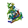
- Structure visualization
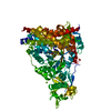
| Structure viewer | Molecule: MolmilJmol/JSmol |
|---|
- Downloads & links
-Download
| PDBx/mmCIF format | 6apz.cif.gz | 158.6 KB | Display | PDBx/mmCIF format |
|---|---|---|---|---|
| PDB format | pdb6apz.ent.gz | 121.1 KB | Display | PDB format |
| PDBx/mmJSON format | 6apz.json.gz | Tree view | PDBx/mmJSON format | |
| Others |  Other downloads Other downloads |
-Validation report
| Arichive directory | https://data.pdbj.org/pub/pdb/validation_reports/ap/6apzftp://data.pdbj.org/pub/pdb/validation_reports/ap/6apz | HTTPS FTP |
|---|
-Related structure data
| Related structure data | 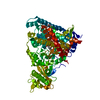 6as6C 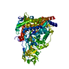 6asuC  6au9C 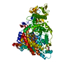 6axbC  6ba7C 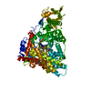 6bu1C 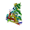 6c2xC 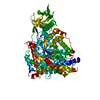 6c6oC 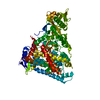 6c7bC  6c8pC 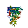 6dkoC 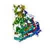 6dl9C 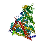 6dljC 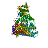 6dnpC  1n8iS S: Starting model for refinement C: citing same article ( |
|---|---|
| Similar structure data |


-Links
 PDBj
PDBj- Assembly
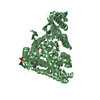
| Deposited unit | 
| ||||||||
|---|---|---|---|---|---|---|---|---|---|
| 1 |
| ||||||||
| Unit cell |
|
-Components
-Protein , 1 types, 1 molecules A
| #1: Protein | Mass: 80456.734 Da / Num. of mol.: 1 / Mutation: C619A Source method: isolated from a genetically manipulated source Source: (gene. exp.) Mycobacterium tuberculosis (strain ATCC 25177 / H37Ra) (bacteria)Strain: ATCC 25177 / H37Ra / Gene: glcB, MRA_1848 / Production host: |
|---|
-Non-polymers , 5 types, 300 molecules 


| #2: Chemical | ChemComp-MG /  Mass: 24.305 Da / Num. of mol.: 1 / Source method: obtained synthetically / Formula: Mg Mass: 24.305 Da / Num. of mol.: 1 / Source method: obtained synthetically / Formula: Mg | ||
|---|---|---|---|
| #3: Chemical | ChemComp-BQ4 / ( Mass: 208.168 Da / Num. of mol.: 1 / Source method: obtained synthetically / Formula: C10H8O5 / Feature type: SUBJECT OF INVESTIGATION Mass: 208.168 Da / Num. of mol.: 1 / Source method: obtained synthetically / Formula: C10H8O5 / Feature type: SUBJECT OF INVESTIGATION | ||
| #4: Chemical | ChemComp-NI /  Mass: 58.693 Da / Num. of mol.: 1 / Source method: isolated from a natural source / Formula: Ni Mass: 58.693 Da / Num. of mol.: 1 / Source method: isolated from a natural source / Formula: Ni | ||
| #5: Chemical | ChemComp-PEG /  Mass: 106.120 Da / Num. of mol.: 4 / Source method: obtained synthetically / Formula: C4H10O3 Mass: 106.120 Da / Num. of mol.: 4 / Source method: obtained synthetically / Formula: C4H10O3#6: Water | ChemComp-HOH / | Mass: 18.015 Da / Num. of mol.: 293 / Source method: isolated from a natural source / Formula: H2O |
-Experimental details
-Experiment
| Experiment | Method: X-RAY DIFFRACTION / Number of used crystals: 1 |
|---|
- Sample preparation
| Crystal | Density Matthews: 2.21 Å3/Da / Density % sol: 44.42 % |
|---|---|
| Crystal grow | Temperature: 289 K / Method: vapor diffusion, hanging drop / Details: MgCl2, TRIS-HCl, PEG3350 / PH range: 7.5-8.5 |
-Data collection
| Diffraction | Mean temperature: 120 K |
|---|---|
| Diffraction source | Source: ROTATING ANODE / Type: RIGAKU MICROMAX-007 / Wavelength: 1.54178 Å |
| Detector | Type: BRUKER SMART 6000 / Detector: CCD / Date: Nov 17, 2011 |
| Radiation | Protocol: SINGLE WAVELENGTH / Monochromatic (M) / Laue (L): M / Scattering type: x-ray |
| Radiation wavelength | Wavelength: 1.54178 Å / Relative weight: 1 |
| Reflection | Resolution: 2.22→50 Å / Num. obs: 26910 / % possible obs: 72.9 % / Redundancy: 12 % / Rmerge(I) obs: 0.118 / Net I/σ(I): 8.2 |
| Reflection shell | Resolution: 2.22→2.26 Å / Rmerge(I) obs: 0.823 |
- Processing
| Software |
| |||||||||||||||||||||||||||||||||||||||||||||||||||||||||||||||||||||||||||||
|---|---|---|---|---|---|---|---|---|---|---|---|---|---|---|---|---|---|---|---|---|---|---|---|---|---|---|---|---|---|---|---|---|---|---|---|---|---|---|---|---|---|---|---|---|---|---|---|---|---|---|---|---|---|---|---|---|---|---|---|---|---|---|---|---|---|---|---|---|---|---|---|---|---|---|---|---|---|---|
| Refinement | Method to determine structure: MOLECULAR REPLACEMENT Starting model: 1N8I Resolution: 2.254→45.022 Å / SU ML: 0.29 / Cross valid method: FREE R-VALUE / σ(F): 1.34 / Phase error: 29.67 / Stereochemistry target values: ML
| |||||||||||||||||||||||||||||||||||||||||||||||||||||||||||||||||||||||||||||
| Solvent computation | Shrinkage radii: 0.9 Å / VDW probe radii: 1.11 Å / Solvent model: FLAT BULK SOLVENT MODEL | |||||||||||||||||||||||||||||||||||||||||||||||||||||||||||||||||||||||||||||
| Refinement step | Cycle: LAST / Resolution: 2.254→45.022 Å
| |||||||||||||||||||||||||||||||||||||||||||||||||||||||||||||||||||||||||||||
| Refine LS restraints |
| |||||||||||||||||||||||||||||||||||||||||||||||||||||||||||||||||||||||||||||
| LS refinement shell |
|
