 Movie
Movie Controller


+ Open data

- Basic information
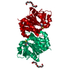
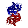
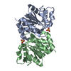
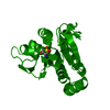
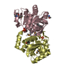
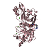
| Entry | Database: PDB / ID: 6af7 | |||||||||||||||
|---|---|---|---|---|---|---|---|---|---|---|---|---|---|---|---|---|
| Title | DJ-1 C106S unbound | |||||||||||||||
 Components Components | Protein/nucleic acid deglycase DJ-1 | |||||||||||||||
 Keywords Keywords | HYDROLASE / DJ-1 / Parkinson's disease / Drug discovery / Fragment-based drug discovery | |||||||||||||||
| Function / homology |  Function and homology information Function and homology informationpositive regulation of acute inflammatory response to antigenic stimulus / tyrosine 3-monooxygenase activator activity / cellular response to glyoxal / L-dopa decarboxylase activator activity / detoxification of hydrogen peroxide / : / : / guanine deglycation, glyoxal removal / cellular detoxification of methylglyoxal / regulation of supramolecular fiber organization ...positive regulation of acute inflammatory response to antigenic stimulus / tyrosine 3-monooxygenase activator activity / cellular response to glyoxal / L-dopa decarboxylase activator activity / detoxification of hydrogen peroxide / : / : / guanine deglycation, glyoxal removal / cellular detoxification of methylglyoxal / regulation of supramolecular fiber organization / negative regulation of death-inducing signaling complex assembly / negative regulation of TRAIL-activated apoptotic signaling pathway / : / glyoxalase (glycolic acid-forming) activity / negative regulation of protein K48-linked deubiquitination / detection of oxidative stress / negative regulation of nitrosative stress-induced intrinsic apoptotic signaling pathway / glycolate biosynthetic process / glyoxal metabolic process / guanine deglycation / detoxification of mercury ion / ubiquitin-protein transferase inhibitor activity / protein deglycase / hydrogen peroxide metabolic process / mercury ion binding / methylglyoxal metabolic process / protein deglycase activity / positive regulation of autophagy of mitochondrion / superoxide dismutase copper chaperone activity / oxidoreductase activity, acting on peroxide as acceptor / positive regulation of dopamine biosynthetic process / positive regulation of mitochondrial electron transport, NADH to ubiquinone / lactate biosynthetic process / negative regulation of hydrogen peroxide-induced neuron intrinsic apoptotic signaling pathway / protein repair / peptidase inhibitor activity / peroxiredoxin activity / cellular detoxification of aldehyde / small protein activating enzyme binding / Hydrolases; Acting on ester bonds; Thioester hydrolases / regulation of oxidative stress-induced neuron intrinsic apoptotic signaling pathway / detoxification of copper ion / negative regulation of protein sumoylation / positive regulation of oxidative stress-induced intrinsic apoptotic signaling pathway / negative regulation of protein export from nucleus / cupric ion binding / negative regulation of oxidative stress-induced neuron intrinsic apoptotic signaling pathway / regulation of androgen receptor signaling pathway / membrane hyperpolarization / Hydrolases; Acting on carbon-nitrogen bonds, other than peptide bonds; In linear amides / oxygen sensor activity / insulin secretion / androgen receptor signaling pathway / nuclear androgen receptor binding / negative regulation of intrinsic apoptotic signaling pathway in response to hydrogen peroxide / ubiquitin-like protein conjugating enzyme binding / ubiquitin-specific protease binding / cytokine binding / positive regulation of reactive oxygen species biosynthetic process / dopamine uptake involved in synaptic transmission / cuprous ion binding / signaling receptor activator activity / regulation of synaptic vesicle endocytosis / single fertilization / membrane depolarization / negative regulation of endoplasmic reticulum stress-induced intrinsic apoptotic signaling pathway / negative regulation of oxidative stress-induced intrinsic apoptotic signaling pathway / negative regulation of reactive oxygen species biosynthetic process / removal of superoxide radicals / SUMOylation of transcription cofactors / negative regulation of protein ubiquitination / negative regulation of proteasomal ubiquitin-dependent protein catabolic process / regulation of neuron apoptotic process / negative regulation of extrinsic apoptotic signaling pathway / positive regulation of interleukin-8 production / regulation of mitochondrial membrane potential / adult locomotory behavior / adherens junction / mitochondrion organization / centriole / positive regulation of protein-containing complex assembly / Late endosomal microautophagy / PML body / mitochondrial intermembrane space / positive regulation of protein localization to nucleus / autophagy / positive regulation of reactive oxygen species metabolic process / enzyme activator activity / kinase binding / cellular response to hydrogen peroxide / Chaperone Mediated Autophagy / Aggrephagy / synaptic vesicle / peptidase activity / glucose homeostasis / cellular response to oxidative stress / cell body / regulation of inflammatory response / response to oxidative stress / scaffold protein binding Similarity search - Function | |||||||||||||||
| Biological species |  Homo sapiens (human) Homo sapiens (human) | |||||||||||||||
| Method |  X-RAY DIFFRACTION / SYNCHROTRON / MOLECULAR REPLACEMENT / Resolution: 1.3 Å X-RAY DIFFRACTION / SYNCHROTRON / MOLECULAR REPLACEMENT / Resolution: 1.3 Å | |||||||||||||||
 Authors Authors | Caaveiro, J.M.M. / Tashiro, S. / Tsumoto, K. | |||||||||||||||
| Funding support |  Japan, 4items Japan, 4items
| |||||||||||||||
 Citation Citation | Journal: ACS Chem. Biol. / Year: 2018 Title: Discovery and Optimization of Inhibitors of the Parkinson's Disease Associated Protein DJ-1. Authors: Tashiro, S. / Caaveiro, J.M.M. / Nakakido, M. / Tanabe, A. / Nagatoishi, S. / Tamura, Y. / Matsuda, N. / Liu, D. / Hoang, Q.Q. / Tsumoto, K. #1: Journal: Biochemistry / Year: 2014Title: Thermodynamic and structural characterization of the specific binding of Zn(II) to human protein DJ-1. Authors: Tashiro, S. / Caaveiro, J.M.M. / Wu, C.X. / Hoang, Q.Q. / Tsumoto, K. | |||||||||||||||
| History |
|
- Structure visualization
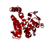
| Structure viewer | Molecule: MolmilJmol/JSmol |
|---|
- Downloads & links
-Download
| PDBx/mmCIF format | 6af7.cif.gz | 100.4 KB | Display | PDBx/mmCIF format |
|---|---|---|---|---|
| PDB format | pdb6af7.ent.gz | 76 KB | Display | PDB format |
| PDBx/mmJSON format | 6af7.json.gz | Tree view | PDBx/mmJSON format | |
| Others |  Other downloads Other downloads |
-Validation report
| Arichive directory | https://data.pdbj.org/pub/pdb/validation_reports/af/6af7ftp://data.pdbj.org/pub/pdb/validation_reports/af/6af7 | HTTPS FTP |
|---|
-Related structure data
| Related structure data |  6af5C  6af9C 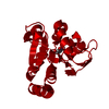 6afaC  6afbC  6afcC  6afdC  6afeC  6affC  6afgC  6afhC  6afiC 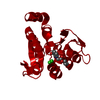 6afjC  6aflC  1soaS S: Starting model for refinement C: citing same article ( |
|---|---|
| Similar structure data |


-Links
 PDBj
PDBj

- Assembly
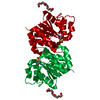
| Deposited unit | 
| ||||||||
|---|---|---|---|---|---|---|---|---|---|
| 1 | 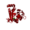
| ||||||||
| Unit cell |
| ||||||||
| Components on special symmetry positions |
|
-Components
| #1: Protein | Mass: 19900.986 Da / Num. of mol.: 1 / Mutation: C106S Source method: isolated from a genetically manipulated source Source: (gene. exp.) Homo sapiens (human) / Gene: PARK7 / Plasmid: pET28B / Production host:  References: UniProt: Q99497, Hydrolases; Acting on ester bonds; Thioester hydrolases, Hydrolases; Acting on carbon-nitrogen bonds, other than peptide bonds; In linear amides, protein deglycase |
|---|---|
| #2: Chemical | ChemComp-1PE /   Mass: 238.278 Da / Num. of mol.: 1 / Source method: obtained synthetically / Formula: C10H22O6 / Comment: precipitant*YM Mass: 238.278 Da / Num. of mol.: 1 / Source method: obtained synthetically / Formula: C10H22O6 / Comment: precipitant*YM |
| #3: Chemical | ChemComp-CL /   Mass: 35.453 Da / Num. of mol.: 1 / Source method: obtained synthetically / Formula: Cl Mass: 35.453 Da / Num. of mol.: 1 / Source method: obtained synthetically / Formula: Cl |
| #4: Water | ChemComp-HOH /  Mass: 18.015 Da / Num. of mol.: 278 / Source method: isolated from a natural source / Formula: H2O Mass: 18.015 Da / Num. of mol.: 278 / Source method: isolated from a natural source / Formula: H2O |
-Experimental details
-Experiment
| Experiment | Method: X-RAY DIFFRACTION / Number of used crystals: 1 |
|---|
- Sample preparation
| Crystal | Density Matthews: 3.06 Å3/Da / Density % sol: 59.76 % |
|---|---|
| Crystal grow | Temperature: 293.15 K / Method: vapor diffusion, hanging drop / pH: 8.5 Details: pH 8.5, 100mM TRIS-HCl, 200mM sodium citrate, 30% PEG 400, 5mM DTT |
-Data collection
| Diffraction | Mean temperature: 100 K |
|---|---|
| Diffraction source | Source: SYNCHROTRON / Site: Photon Factory / Beamline: BL-5A / Wavelength: 1 Å |
| Detector | Type: ADSC QUANTUM 315 / Detector: CCD / Date: Apr 17, 2013 |
| Radiation | Protocol: SINGLE WAVELENGTH / Monochromatic (M) / Laue (L): M / Scattering type: x-ray |
| Radiation wavelength | Wavelength: 1 Å / Relative weight: 1 |
| Reflection | Resolution: 1.3→26.6 Å / Num. obs: 60285 / % possible obs: 100 % / Redundancy: 5.3 % / Rmerge(I) obs: 0.103 / Net I/σ(I): 10.1 |
| Reflection shell | Resolution: 1.3→1.37 Å / Redundancy: 5.3 % / Rmerge(I) obs: 0.354 / Mean I/σ(I) obs: 4.2 / % possible all: 100 |
- Processing
| Software |
| ||||||||||||||||||||||||||||||||||||||||||||||||||||||||||||||||||||||||||||||||||||||||||||||||||||||||||||||||||||||||||||||||||||||||||||||||||||||||||||||||||||||||||||||||||||||
|---|---|---|---|---|---|---|---|---|---|---|---|---|---|---|---|---|---|---|---|---|---|---|---|---|---|---|---|---|---|---|---|---|---|---|---|---|---|---|---|---|---|---|---|---|---|---|---|---|---|---|---|---|---|---|---|---|---|---|---|---|---|---|---|---|---|---|---|---|---|---|---|---|---|---|---|---|---|---|---|---|---|---|---|---|---|---|---|---|---|---|---|---|---|---|---|---|---|---|---|---|---|---|---|---|---|---|---|---|---|---|---|---|---|---|---|---|---|---|---|---|---|---|---|---|---|---|---|---|---|---|---|---|---|---|---|---|---|---|---|---|---|---|---|---|---|---|---|---|---|---|---|---|---|---|---|---|---|---|---|---|---|---|---|---|---|---|---|---|---|---|---|---|---|---|---|---|---|---|---|---|---|---|---|
| Refinement | Method to determine structure: MOLECULAR REPLACEMENT Starting model: 1SOA Resolution: 1.3→26.6 Å / Cor.coef. Fo:Fc: 0.984 / Cor.coef. Fo:Fc free: 0.981 / SU B: 0.858 / SU ML: 0.016 / Cross valid method: THROUGHOUT / ESU R: 0.027 / ESU R Free: 0.028 / Details: HYDROGENS HAVE BEEN ADDED IN THE RIDING POSITIONS
| ||||||||||||||||||||||||||||||||||||||||||||||||||||||||||||||||||||||||||||||||||||||||||||||||||||||||||||||||||||||||||||||||||||||||||||||||||||||||||||||||||||||||||||||||||||||
| Solvent computation | Ion probe radii: 0.8 Å / Shrinkage radii: 0.8 Å / VDW probe radii: 1.2 Å | ||||||||||||||||||||||||||||||||||||||||||||||||||||||||||||||||||||||||||||||||||||||||||||||||||||||||||||||||||||||||||||||||||||||||||||||||||||||||||||||||||||||||||||||||||||||
| Displacement parameters | Biso mean: 15.515 Å2
| ||||||||||||||||||||||||||||||||||||||||||||||||||||||||||||||||||||||||||||||||||||||||||||||||||||||||||||||||||||||||||||||||||||||||||||||||||||||||||||||||||||||||||||||||||||||
| Refinement step | Cycle: 1 / Resolution: 1.3→26.6 Å
| ||||||||||||||||||||||||||||||||||||||||||||||||||||||||||||||||||||||||||||||||||||||||||||||||||||||||||||||||||||||||||||||||||||||||||||||||||||||||||||||||||||||||||||||||||||||
| Refine LS restraints |
|