 Movie
Movie Controller

[English] 日本語
 Yorodumi
- PDB-6a4j: Crystal structure of Thioredoxin reductase 2 from Staphylococcus ... -
+ Open data

- Basic information
| Entry | Database: PDB / ID: 6a4j | |||||||||
|---|---|---|---|---|---|---|---|---|---|---|
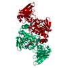
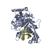
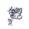
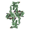
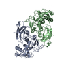
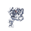
| Title | Crystal structure of Thioredoxin reductase 2 from Staphylococcus aureus | |||||||||
 Components Components | Ferredoxin--NADP reductase | |||||||||
 Keywords Keywords | OXIDOREDUCTASE / flavoenzyme | |||||||||
| Function / homology |  Function and homology information Function and homology informationferredoxin-NADP+ reductase / ferredoxin-NADP+ reductase activity / thioredoxin-disulfide reductase (NADPH) activity / cell redox homeostasis / monooxygenase activity / NADP binding / flavin adenine dinucleotide binding Similarity search - Function | |||||||||
| Biological species |   Staphylococcus aureus (bacteria) Staphylococcus aureus (bacteria) | |||||||||
| Method |  X-RAY DIFFRACTION / MOLECULAR REPLACEMENT / Resolution: 3.4 Å X-RAY DIFFRACTION / MOLECULAR REPLACEMENT / Resolution: 3.4 Å | |||||||||
 Authors Authors | Bose, M. / Bhattacharyya, S. / Ghosh, A.K. / Das, A.K. | |||||||||
| Funding support |  India, 1items India, 1items
| |||||||||
 Citation Citation | Journal: Biochimie / Year: 2019 Title: Elucidation of the mechanism of disulfide exchange between staphylococcal thioredoxin2 and thioredoxin reductase2: A structural insight. Authors: Bose, M. / Bhattacharyya, S. / Biswas, R. / Roychowdhury, A. / Bhattacharjee, A. / Ghosh, A.K. / Das, A.K. | |||||||||
| History |
|
- Structure visualization
| Structure viewer | Molecule: MolmilJmol/JSmol |
|---|
- Downloads & links
-Download
| PDBx/mmCIF format | 6a4j.cif.gz | 268.4 KB | Display | PDBx/mmCIF format |
|---|---|---|---|---|
| PDB format | pdb6a4j.ent.gz | 215.6 KB | Display | PDB format |
| PDBx/mmJSON format | 6a4j.json.gz | Tree view | PDBx/mmJSON format | |
| Others |  Other downloads Other downloads |
-Validation report
| Arichive directory | https://data.pdbj.org/pub/pdb/validation_reports/a4/6a4jftp://data.pdbj.org/pub/pdb/validation_reports/a4/6a4j | HTTPS FTP |
|---|
-Related structure data
| Related structure data |  4ruvC  5twcS S: Starting model for refinement C: citing same article ( |
|---|---|
| Similar structure data |


-Links
 PDBj
PDBj


- Assembly
| Deposited unit | 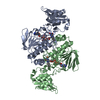
| ||||||||
|---|---|---|---|---|---|---|---|---|---|
| 1 |
| ||||||||
| Unit cell |
|
-Components
| #1: Protein | Mass: 38288.531 Da / Num. of mol.: 2 Source method: isolated from a genetically manipulated source Source: (gene. exp.) Staphylococcus aureus (bacteria)Gene: trxB_2, EP54_04310, EQ90_00075, RK60_12235, RK64_12650, RK68_02995, RK73_09520, RK98_08250, RL06_05030, SAMEA3448991_00661 Plasmid: pQE30 / Production host: References: UniProt: A0A1Q4GXS1, UniProt: Q2FVP8*PLUS, ferredoxin-NADP+ reductase #2: Chemical |   Mass: 785.550 Da / Num. of mol.: 2 Mass: 785.550 Da / Num. of mol.: 2Source method: isolated from a genetically manipulated source Formula: C27H33N9O15P2 / Comment: FAD*YM Has protein modification | Y | |
|---|
-Experimental details
-Experiment
| Experiment | Method: X-RAY DIFFRACTION / Number of used crystals: 1 |
|---|
- Sample preparation
| Crystal | Density Matthews: 3.3 Å3/Da / Density % sol: 62.74 % |
|---|---|
| Crystal grow | Temperature: 298 K / Method: vapor diffusion, hanging drop / pH: 7 / Details: 2.4 M sodium malonate pH 7.0 |
-Data collection
| Diffraction | Mean temperature: 100 K |
|---|---|
| Diffraction source | Source: ROTATING ANODE / Type: RIGAKU MICROMAX-007 / Wavelength: 1.5418 Å |
| Detector | Type: RIGAKU RAXIS IV++ / Detector: IMAGE PLATE / Date: Nov 28, 2017 / Details: varimax osmic mirror |
| Radiation | Protocol: SINGLE WAVELENGTH / Monochromatic (M) / Laue (L): M / Scattering type: x-ray |
| Radiation wavelength | Wavelength: 1.5418 Å / Relative weight: 1 |
| Reflection | Resolution: 3.4→19.861 Å / Num. obs: 14419 / % possible obs: 99.3 % / Redundancy: 2 % / CC1/2: 0.979 / Rmerge(I) obs: 0.1193 / Rpim(I) all: 0.1193 / Rrim(I) all: 0.1687 / Net I/σ(I): 6.01 |
| Reflection shell | Resolution: 3.4→19.86 Å / Rmerge(I) obs: 0.3953 / Num. unique obs: 1427 / CC1/2: 0.658 / Rpim(I) all: 0.3953 / Rrim(I) all: 0.559 |
- Processing
| Software |
| ||||||||||||||||||||||||||||||||||||||||||
|---|---|---|---|---|---|---|---|---|---|---|---|---|---|---|---|---|---|---|---|---|---|---|---|---|---|---|---|---|---|---|---|---|---|---|---|---|---|---|---|---|---|---|---|
| Refinement | Method to determine structure: MOLECULAR REPLACEMENT Starting model: 5TWC Resolution: 3.4→19.861 Å / SU ML: 0.28 / Cross valid method: FREE R-VALUE / σ(F): 1.34 / Phase error: 24.56
| ||||||||||||||||||||||||||||||||||||||||||
| Solvent computation | Shrinkage radii: 0.9 Å / VDW probe radii: 1.11 Å | ||||||||||||||||||||||||||||||||||||||||||
| Refinement step | Cycle: LAST / Resolution: 3.4→19.861 Å
| ||||||||||||||||||||||||||||||||||||||||||
| Refine LS restraints |
| ||||||||||||||||||||||||||||||||||||||||||
| LS refinement shell |
| ||||||||||||||||||||||||||||||||||||||||||
| Refinement TLS params. | Method: refined / Origin x: -4.6846 Å / Origin y: 16.8835 Å / Origin z: -53.5689 Å
| ||||||||||||||||||||||||||||||||||||||||||
| Refinement TLS group | Selection details: all |
