 Movie
Movie Controller


+ Open data

- Basic information
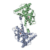
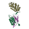
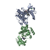
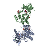
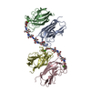
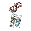
| Entry | Database: PDB / ID: 6a28 | ||||||||||||
|---|---|---|---|---|---|---|---|---|---|---|---|---|---|
| Title | Crystal structure of PprA W183R mutant form 2 | ||||||||||||
 Components Components | DNA repair protein PprA | ||||||||||||
 Keywords Keywords | DNA BINDING PROTEIN | ||||||||||||
| Function / homology | cellular response to desiccation / cellular response to gamma radiation / double-strand break repair via nonhomologous end joining / double-stranded DNA binding / damaged DNA binding / DNA repair / DNA repair protein PprA Function and homology information Function and homology information | ||||||||||||
| Biological species |  Deinococcus radiodurans (radioresistant) Deinococcus radiodurans (radioresistant) | ||||||||||||
| Method |  X-RAY DIFFRACTION / SYNCHROTRON / FOURIER SYNTHESIS / Resolution: 2.193 Å X-RAY DIFFRACTION / SYNCHROTRON / FOURIER SYNTHESIS / Resolution: 2.193 Å | ||||||||||||
 Authors Authors | Adachi, M. / Shibazaki, C. / Shimizu, R. / Arai, S. / Satoh, K. / Narumi, I. / Kuroki, R. | ||||||||||||
| Funding support |  Japan, 3items Japan, 3items
| ||||||||||||
 Citation Citation | Journal: FASEB J. / Year: 2019 Title: Extended structure of pleiotropic DNA repair-promoting protein PprA from Deinococcus radiodurans. Authors: Adachi, M. / Shimizu, R. / Shibazaki, C. / Satoh, K. / Fujiwara, S. / Arai, S. / Narumi, I. / Kuroki, R. | ||||||||||||
| History |
|
- Structure visualization
| Structure viewer | Molecule: MolmilJmol/JSmol |
|---|
- Downloads & links
-Download
| PDBx/mmCIF format | 6a28.cif.gz | 107.2 KB | Display | PDBx/mmCIF format |
|---|---|---|---|---|
| PDB format | pdb6a28.ent.gz | 81.2 KB | Display | PDB format |
| PDBx/mmJSON format | 6a28.json.gz | Tree view | PDBx/mmJSON format | |
| Others |  Other downloads Other downloads |
-Validation report
| Summary document | 6a28_validation.pdf.gz | 450.1 KB | Display | wwPDB validaton report |
|---|---|---|---|---|
| Full document | 6a28_full_validation.pdf.gz | 455.9 KB | Display | |
| Data in XML | 6a28_validation.xml.gz | 20.4 KB | Display | |
| Data in CIF | 6a28_validation.cif.gz | 29.3 KB | Display | |
| Arichive directory | https://data.pdbj.org/pub/pdb/validation_reports/a2/6a28ftp://data.pdbj.org/pub/pdb/validation_reports/a2/6a28 | HTTPS FTP |
-Related structure data
| Related structure data |  6a27C 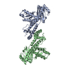 6a29SC S: Starting model for refinement C: citing same article ( |
|---|---|
| Similar structure data |

-Links
 PDBj
PDBj- Assembly
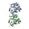
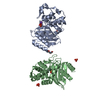
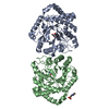
| Deposited unit | 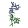
| ||||||||
|---|---|---|---|---|---|---|---|---|---|
| 1 |
| ||||||||
| Unit cell |
|
-Components
| #1: Protein | Mass: 30783.566 Da / Num. of mol.: 2 / Mutation: W183R Source method: isolated from a genetically manipulated source Source: (gene. exp.) Deinococcus radiodurans (radioresistant)Strain: ATCC 13939 / DSM 20539 / JCM 16871 / LMG 4051 / NBRC 15346 / NCIMB 9279 / R1 / VKM B-1422 Gene: pprA, DR_A0346 / Production host: #2: Chemical | ChemComp-SO4 / |   Mass: 96.063 Da / Num. of mol.: 1 / Source method: isolated from a natural source / Formula: SO4 Mass: 96.063 Da / Num. of mol.: 1 / Source method: isolated from a natural source / Formula: SO4#3: Water | ChemComp-HOH / |  Mass: 18.015 Da / Num. of mol.: 232 / Source method: isolated from a natural source / Formula: H2O Mass: 18.015 Da / Num. of mol.: 232 / Source method: isolated from a natural source / Formula: H2OHas protein modification | Y | |
|---|
-Experimental details
-Experiment
| Experiment | Method: X-RAY DIFFRACTION / Number of used crystals: 1 |
|---|
- Sample preparation
| Crystal | Density Matthews: 2.49 Å3/Da / Density % sol: 50.55 % |
|---|---|
| Crystal grow | Temperature: 293 K / Method: vapor diffusion, hanging drop / pH: 8.5 Details: 0.1 M Tris buffer (pH 8.5) containing 0.2 M LiSO4 and 30% PEG4000 |
-Data collection
| Diffraction | Mean temperature: 100 K |
|---|---|
| Diffraction source | Source: SYNCHROTRON / Site: Photon Factory / Beamline: AR-NE3A / Wavelength: 0.97965 Å |
| Detector | Type: ADSC QUANTUM 270 / Detector: CCD / Date: Dec 17, 2009 |
| Radiation | Protocol: SINGLE WAVELENGTH / Monochromatic (M) / Laue (L): M / Scattering type: x-ray |
| Radiation wavelength | Wavelength: 0.97965 Å / Relative weight: 1 |
| Reflection | Resolution: 2.193→46.9 Å / Num. obs: 30255 / % possible obs: 94.8 % / Redundancy: 11.1 % / Rmerge(I) obs: 0.096 / Net I/σ(I): 36.9 |
| Reflection shell | Resolution: 2.2→2.24 Å / Redundancy: 8.2 % / Rmerge(I) obs: 0.513 / Mean I/σ(I) obs: 3.6 / Num. unique obs: 1496 / % possible all: 92.3 |
- Processing
| Software |
| ||||||||||||||||||||||||||||||||||||||||||||||||||||||||||||||||||||||||||||||||||||
|---|---|---|---|---|---|---|---|---|---|---|---|---|---|---|---|---|---|---|---|---|---|---|---|---|---|---|---|---|---|---|---|---|---|---|---|---|---|---|---|---|---|---|---|---|---|---|---|---|---|---|---|---|---|---|---|---|---|---|---|---|---|---|---|---|---|---|---|---|---|---|---|---|---|---|---|---|---|---|---|---|---|---|---|---|---|
| Refinement | Method to determine structure: FOURIER SYNTHESIS Starting model: 6A29 Resolution: 2.193→32.108 Å / SU ML: 0.24 / Cross valid method: FREE R-VALUE / σ(F): 1.34 / Phase error: 23.14
| ||||||||||||||||||||||||||||||||||||||||||||||||||||||||||||||||||||||||||||||||||||
| Solvent computation | Shrinkage radii: 0.9 Å / VDW probe radii: 1.11 Å | ||||||||||||||||||||||||||||||||||||||||||||||||||||||||||||||||||||||||||||||||||||
| Refinement step | Cycle: LAST / Resolution: 2.193→32.108 Å
| ||||||||||||||||||||||||||||||||||||||||||||||||||||||||||||||||||||||||||||||||||||
| Refine LS restraints |
| ||||||||||||||||||||||||||||||||||||||||||||||||||||||||||||||||||||||||||||||||||||
| LS refinement shell |
|
