 Movie
Movie Controller

[English] 日本語
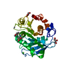
 Yorodumi
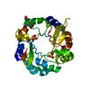
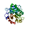
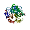
- PDB-5zno: Crystal structure of PET-degrading cutinase Cut190 S176A/S226P/R2... -
+ Open data

- Basic information
| Entry | Database: PDB / ID: 5zno | ||||||
|---|---|---|---|---|---|---|---|
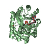
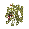
| Title | Crystal structure of PET-degrading cutinase Cut190 S176A/S226P/R228S/ mutant in Ca(2+)-bound state | ||||||
 Components Components | Alpha/beta hydrolase family protein | ||||||
 Keywords Keywords | HYDROLASE / POLYESTERASE / ALPHA/BETA-HYDROLASE FOLD / PROTEIN ENGINEERING / THERMOSTABILITY | ||||||
| Function / homology |  Function and homology information Function and homology information | ||||||
| Biological species |  Saccharomonospora viridis (bacteria) Saccharomonospora viridis (bacteria) | ||||||
| Method |  X-RAY DIFFRACTION / SYNCHROTRON / MOLECULAR REPLACEMENT / Resolution: 1.60264346898 Å X-RAY DIFFRACTION / SYNCHROTRON / MOLECULAR REPLACEMENT / Resolution: 1.60264346898 Å | ||||||
 Authors Authors | Numoto, N. / Inaba, S. / Yamagami, Y. / Kamiya, N. / Bekker, G.J. / Ishii, K. / Uchiyama, S. / Kawai, F. / Ito, N. / Oda, M. | ||||||
 Citation Citation | Journal: Biochemistry / Year: 2018 Title: Structural Dynamics of the PET-Degrading Cutinase-like Enzyme from Saccharomonospora viridis AHK190 in Substrate-Bound States Elucidates the Ca2+-Driven Catalytic Cycle. Authors: Numoto, N. / Kamiya, N. / Bekker, G.J. / Yamagami, Y. / Inaba, S. / Ishii, K. / Uchiyama, S. / Kawai, F. / Ito, N. / Oda, M. | ||||||
| History |
|
- Structure visualization
| Structure viewer | Molecule: MolmilJmol/JSmol |
|---|
- Downloads & links
-Download
| PDBx/mmCIF format | 5zno.cif.gz | 166.9 KB | Display | PDBx/mmCIF format |
|---|---|---|---|---|
| PDB format | pdb5zno.ent.gz | 104.2 KB | Display | PDB format |
| PDBx/mmJSON format | 5zno.json.gz | Tree view | PDBx/mmJSON format | |
| Others |  Other downloads Other downloads |
-Validation report
| Arichive directory | https://data.pdbj.org/pub/pdb/validation_reports/zn/5znoftp://data.pdbj.org/pub/pdb/validation_reports/zn/5zno | HTTPS FTP |
|---|
-Related structure data
| Related structure data |  5zrqC  5zrrC  5zrsC 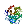 4wfjS S: Starting model for refinement C: citing same article ( |
|---|---|
| Similar structure data |

-Links
 PDBj
PDBj- Assembly
| Deposited unit | 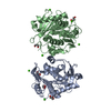
| ||||||||||||
|---|---|---|---|---|---|---|---|---|---|---|---|---|---|
| 1 | 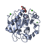
| ||||||||||||
| 2 | 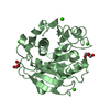
| ||||||||||||
| Unit cell |
|
-Components
| #1: Protein | Mass: 29193.658 Da / Num. of mol.: 2 / Mutation: S176A, S226P, R228S Source method: isolated from a genetically manipulated source Source: (gene. exp.) Saccharomonospora viridis (bacteria) / Strain: AHK190 / Gene: Cut190, SAMN02982918_2340 / Plasmid: pQE80L / Production host: #2: Chemical | ChemComp-CA /   Mass: 40.078 Da / Num. of mol.: 7 / Source method: obtained synthetically / Formula: Ca Mass: 40.078 Da / Num. of mol.: 7 / Source method: obtained synthetically / Formula: Ca#3: Chemical | ChemComp-GOL /   Mass: 92.094 Da / Num. of mol.: 5 / Source method: obtained synthetically / Formula: C3H8O3 Mass: 92.094 Da / Num. of mol.: 5 / Source method: obtained synthetically / Formula: C3H8O3#4: Water | ChemComp-HOH / |  Mass: 18.015 Da / Num. of mol.: 789 / Source method: isolated from a natural source / Formula: H2O Mass: 18.015 Da / Num. of mol.: 789 / Source method: isolated from a natural source / Formula: H2OHas protein modification | Y | |
|---|
-Experimental details
-Experiment
| Experiment | Method: X-RAY DIFFRACTION / Number of used crystals: 1 |
|---|
- Sample preparation
| Crystal | Density Matthews: 2.52 Å3/Da / Density % sol: 51.26 % |
|---|---|
| Crystal grow | Temperature: 293 K / Method: vapor diffusion, hanging drop / pH: 7.5 Details: 0.1 M HEPES pH 7.5, 10% w/v PEG 8000, 8% v/v Ethylene glycol |
-Data collection
| Diffraction | Mean temperature: 95 K |
|---|---|
| Diffraction source | Source: SYNCHROTRON / Site: Photon Factory  / Beamline: BL-1A / Wavelength: 1.1 Å / Beamline: BL-1A / Wavelength: 1.1 Å |
| Detector | Type: DECTRIS EIGER X 4M / Detector: PIXEL / Date: Dec 9, 2016 |
| Radiation | Monochromator: Cryo-cooled channel-cut Si(111) / Protocol: SINGLE WAVELENGTH / Monochromatic (M) / Laue (L): M / Scattering type: x-ray |
| Radiation wavelength | Wavelength: 1.1 Å / Relative weight: 1 |
| Reflection | Resolution: 1.6→50 Å / Num. obs: 75063 / % possible obs: 99.3 % / Redundancy: 3.3 % / Biso Wilson estimate: 15.4807795741 Å2 / CC1/2: 0.992 / Rsym value: 0.136 / Net I/σ(I): 6.8 |
| Reflection shell | Resolution: 1.6→1.7 Å / Redundancy: 3.2 % / Num. unique obs: 12065 / CC1/2: 0.518 / Rsym value: 0.95 / % possible all: 99.3 |
- Processing
| Software |
| ||||||||||||||||||||||||||||||||||||||||||||||||||||||||||||||||||||||||||||||||||||||||||||||||||||||||||||||||||||||||||||||||||||||||||||||||||||||||||||||||||||||||||||||||||||||||||||||||||||
|---|---|---|---|---|---|---|---|---|---|---|---|---|---|---|---|---|---|---|---|---|---|---|---|---|---|---|---|---|---|---|---|---|---|---|---|---|---|---|---|---|---|---|---|---|---|---|---|---|---|---|---|---|---|---|---|---|---|---|---|---|---|---|---|---|---|---|---|---|---|---|---|---|---|---|---|---|---|---|---|---|---|---|---|---|---|---|---|---|---|---|---|---|---|---|---|---|---|---|---|---|---|---|---|---|---|---|---|---|---|---|---|---|---|---|---|---|---|---|---|---|---|---|---|---|---|---|---|---|---|---|---|---|---|---|---|---|---|---|---|---|---|---|---|---|---|---|---|---|---|---|---|---|---|---|---|---|---|---|---|---|---|---|---|---|---|---|---|---|---|---|---|---|---|---|---|---|---|---|---|---|---|---|---|---|---|---|---|---|---|---|---|---|---|---|---|---|---|
| Refinement | Method to determine structure: MOLECULAR REPLACEMENT Starting model: 4WFJ Resolution: 1.60264346898→47.7178508489 Å / SU ML: 0.235868861104 / Cross valid method: FREE R-VALUE / σ(F): 1.3474730099 / Phase error: 22.9434927349
| ||||||||||||||||||||||||||||||||||||||||||||||||||||||||||||||||||||||||||||||||||||||||||||||||||||||||||||||||||||||||||||||||||||||||||||||||||||||||||||||||||||||||||||||||||||||||||||||||||||
| Solvent computation | Shrinkage radii: 0.9 Å / VDW probe radii: 1.11 Å | ||||||||||||||||||||||||||||||||||||||||||||||||||||||||||||||||||||||||||||||||||||||||||||||||||||||||||||||||||||||||||||||||||||||||||||||||||||||||||||||||||||||||||||||||||||||||||||||||||||
| Displacement parameters | Biso mean: 18.919072451 Å2 | ||||||||||||||||||||||||||||||||||||||||||||||||||||||||||||||||||||||||||||||||||||||||||||||||||||||||||||||||||||||||||||||||||||||||||||||||||||||||||||||||||||||||||||||||||||||||||||||||||||
| Refinement step | Cycle: LAST / Resolution: 1.60264346898→47.7178508489 Å
| ||||||||||||||||||||||||||||||||||||||||||||||||||||||||||||||||||||||||||||||||||||||||||||||||||||||||||||||||||||||||||||||||||||||||||||||||||||||||||||||||||||||||||||||||||||||||||||||||||||
| Refine LS restraints |
| ||||||||||||||||||||||||||||||||||||||||||||||||||||||||||||||||||||||||||||||||||||||||||||||||||||||||||||||||||||||||||||||||||||||||||||||||||||||||||||||||||||||||||||||||||||||||||||||||||||
| LS refinement shell |
|
