+ Open data
- Basic information
| Entry | Database: PDB / ID: 5zmp | ||||||
|---|---|---|---|---|---|---|---|
| Title | The structure of a lysine deacylase | ||||||
 Components Components | lysine deacylase | ||||||
 Keywords Keywords | HYDROLASE / lysine deacylase | ||||||
| Function / homology | : / Histone deacetylase family / Histone deacetylase domain / Histone deacetylase domain superfamily / Histone deacetylase domain / Ureohydrolase domain superfamily / hydrolase activity / metal ion binding / Acetylpolyamine aminohydrolase Function and homology information Function and homology information | ||||||
| Biological species |  Synechococcus sp. PCC 73109 (bacteria) Synechococcus sp. PCC 73109 (bacteria) | ||||||
| Method |  X-RAY DIFFRACTION / X-RAY DIFFRACTION /  SYNCHROTRON / SYNCHROTRON /  MOLECULAR REPLACEMENT / Resolution: 2.19 Å MOLECULAR REPLACEMENT / Resolution: 2.19 Å | ||||||
 Authors Authors | Ge, F. | ||||||
 Citation Citation | |||||||
| History |
|
- Structure visualization
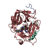
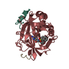
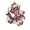
| Structure viewer | Molecule:  Molmil Molmil Jmol/JSmol Jmol/JSmol |
|---|
- Downloads & links
- Download
| PDBx/mmCIF format |  5zmp.cif.gz 5zmp.cif.gz | 78.5 KB | Display |  PDBx/mmCIF format PDBx/mmCIF format |
|---|---|---|---|---|
| PDB format |  pdb5zmp.ent.gz pdb5zmp.ent.gz | 56.8 KB | Display |  PDB format PDB format |
| PDBx/mmJSON format |  5zmp.json.gz 5zmp.json.gz | Tree view |  PDBx/mmJSON format PDBx/mmJSON format | |
| Others |  Other downloads Other downloads |
-Validation report
| Summary document |  5zmp_validation.pdf.gz 5zmp_validation.pdf.gz | 425.8 KB | Display |  wwPDB validaton report wwPDB validaton report |
|---|---|---|---|---|
| Full document |  5zmp_full_validation.pdf.gz 5zmp_full_validation.pdf.gz | 426.5 KB | Display | |
| Data in XML |  5zmp_validation.xml.gz 5zmp_validation.xml.gz | 14.7 KB | Display | |
| Data in CIF |  5zmp_validation.cif.gz 5zmp_validation.cif.gz | 21.4 KB | Display | |
| Arichive directory |  https://data.pdbj.org/pub/pdb/validation_reports/zm/5zmp https://data.pdbj.org/pub/pdb/validation_reports/zm/5zmp ftp://data.pdbj.org/pub/pdb/validation_reports/zm/5zmp ftp://data.pdbj.org/pub/pdb/validation_reports/zm/5zmp | HTTPS FTP |
-Related structure data
| Related structure data |  5g11S S: Starting model for refinement |
|---|---|
| Similar structure data |
- Links
Links
- Assembly
| Deposited unit | 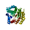
| ||||||||
|---|---|---|---|---|---|---|---|---|---|
| 1 |
| ||||||||
| Unit cell |
|
- Components
| #1: Protein | Mass: 34090.574 Da / Num. of mol.: 1 Source method: isolated from a genetically manipulated source Source: (gene. exp.)  Synechococcus sp. PCC 73109 (bacteria) / Gene: SYNPCC7002_A2791 / Production host: Synechococcus sp. PCC 73109 (bacteria) / Gene: SYNPCC7002_A2791 / Production host:  |
|---|---|
| #2: Chemical | ChemComp-ZN / |
| #3: Water | ChemComp-HOH / |
| Source details | Protein used in this structure is derived from Tax ID 374982 (Synechococcus sp. PCC 73109). ...Protein used in this structure is derived from Tax ID 374982 (Synechococcus sp. PCC 73109). Reference source is Tax ID 32049 (Synechococcus sp. PCC 7002). Sequences are 100 % matched. |
-Experimental details
-Experiment
| Experiment | Method:  X-RAY DIFFRACTION / Number of used crystals: 1 X-RAY DIFFRACTION / Number of used crystals: 1 |
|---|
- Sample preparation
| Crystal | Density Matthews: 2.66 Å3/Da / Density % sol: 53.78 % |
|---|---|
| Crystal grow | Temperature: 291 K / Method: vapor diffusion, hanging drop Details: 1.26 M Sodium phosphate monobasic monohydrate, 0.14 M Potassium phosphate dibasic |
-Data collection
| Diffraction | Mean temperature: 100 K / Serial crystal experiment: N |
|---|---|
| Diffraction source | Source:  SYNCHROTRON / Site: SYNCHROTRON / Site:  SSRF SSRF  / Beamline: BL17U1 / Wavelength: 0.979 Å / Beamline: BL17U1 / Wavelength: 0.979 Å |
| Detector | Type: DECTRIS PILATUS 6M / Detector: PIXEL / Date: Mar 20, 2018 |
| Radiation | Protocol: SINGLE WAVELENGTH / Monochromatic (M) / Laue (L): M / Scattering type: x-ray |
| Radiation wavelength | Wavelength: 0.979 Å / Relative weight: 1 |
| Reflection | Resolution: 2.19→50.7 Å / Num. obs: 18344 / % possible obs: 99.8 % / Redundancy: 7 % / Rmerge(I) obs: 0.214 / Rpim(I) all: 0.132 / Net I/σ(I): 8.7 |
| Reflection shell | Resolution: 2.19→2.25 Å / Redundancy: 7.2 % / Rmerge(I) obs: 1.13 / Mean I/σ(I) obs: 2.2 / Num. unique obs: 1342 / Rpim(I) all: 0.681 / % possible all: 99.8 |
- Processing
| Software |
| ||||||||||||||||
|---|---|---|---|---|---|---|---|---|---|---|---|---|---|---|---|---|---|
| Refinement | Method to determine structure:  MOLECULAR REPLACEMENT MOLECULAR REPLACEMENTStarting model: 5G11 Resolution: 2.19→50 Å / Cross valid method: FREE R-VALUE
| ||||||||||||||||
| Refinement step | Cycle: LAST / Resolution: 2.19→50 Å
| ||||||||||||||||
| LS refinement shell | Resolution: 2.19→2.25 Å /
|
 Movie
Movie Controller






 PDBj
PDBj


