 Movie
Movie Controller

[English] 日本語
 Yorodumi
- PDB-5zkk: Crystal structure of Phosphoserine phosphatase from Entamoeba his... -
+ Open data

- Basic information
| Entry | Database: PDB / ID: 5zkk | ||||||
|---|---|---|---|---|---|---|---|
| Title | Crystal structure of Phosphoserine phosphatase from Entamoeba histolytica | ||||||
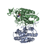
 Components Components | Phosphoglycerate mutase family protein, putative | ||||||
 Keywords Keywords | HYDROLASE / Histidine phosphatase superfamily memeber / Phosphatase / cysteine biosynthetic pathway / BIOSYNTHETIC PROTEIN | ||||||
| Function / homology |  Function and homology information Function and homology information | ||||||
| Biological species |  Entamoeba histolytica HM-1:IMSS-A (eukaryote) Entamoeba histolytica HM-1:IMSS-A (eukaryote) | ||||||
| Method |  X-RAY DIFFRACTION / MOLECULAR REPLACEMENT / Resolution: 2 Å X-RAY DIFFRACTION / MOLECULAR REPLACEMENT / Resolution: 2 Å | ||||||
 Authors Authors | Kumari, P. / Gourinath, S. | ||||||
 Citation Citation | Journal: J.Struct.Biol. / Year: 2019 Title: Structural and functional characterisation of phosphoserine phosphatase, that plays critical role in the oxidative stress response in the parasite Entamoeba histolytica. Authors: Kumari, P. / Babuta, M. / Bhattacharya, A. / Gourinath, S. | ||||||
| History |
|
- Structure visualization
| Structure viewer | Molecule: MolmilJmol/JSmol |
|---|
- Downloads & links
-Download
| PDBx/mmCIF format | 5zkk.cif.gz | 99.3 KB | Display | PDBx/mmCIF format |
|---|---|---|---|---|
| PDB format | pdb5zkk.ent.gz | 74.2 KB | Display | PDB format |
| PDBx/mmJSON format | 5zkk.json.gz | Tree view | PDBx/mmJSON format | |
| Others |  Other downloads Other downloads |
-Validation report
| Arichive directory | https://data.pdbj.org/pub/pdb/validation_reports/zk/5zkkftp://data.pdbj.org/pub/pdb/validation_reports/zk/5zkk | HTTPS FTP |
|---|
-Related structure data
| Related structure data |  5zr2C 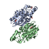 4ij5S S: Starting model for refinement C: citing same article ( |
|---|---|
| Similar structure data |


-Links
 PDBj
PDBj


- Assembly
| Deposited unit | 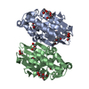
| ||||||||
|---|---|---|---|---|---|---|---|---|---|
| 1 |
| ||||||||
| Unit cell |
|
-Components
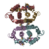
-Protein , 1 types, 2 molecules AB
| #1: Protein | Mass: 23771.080 Da / Num. of mol.: 2 Source method: isolated from a genetically manipulated source Source: (gene. exp.) Entamoeba histolytica HM-1:IMSS-A (eukaryote)Gene: EHI7A_041450 / Plasmid: pET 28b / Production host:  |
|---|
-Non-polymers , 7 types, 229 molecules 


| #2: Chemical |  Mass: 78.133 Da / Num. of mol.: 2 / Source method: obtained synthetically / Formula: C2H6OS Mass: 78.133 Da / Num. of mol.: 2 / Source method: obtained synthetically / Formula: C2H6OS#3: Chemical |  Mass: 94.971 Da / Num. of mol.: 2 / Source method: obtained synthetically / Formula: PO4 Mass: 94.971 Da / Num. of mol.: 2 / Source method: obtained synthetically / Formula: PO4#4: Chemical | ChemComp-PEG /  Mass: 106.120 Da / Num. of mol.: 5 / Source method: obtained synthetically / Formula: C4H10O3 Mass: 106.120 Da / Num. of mol.: 5 / Source method: obtained synthetically / Formula: C4H10O3#5: Chemical | ChemComp-144 / |  Mass: 122.143 Da / Num. of mol.: 1 / Source method: obtained synthetically / Formula: C4H12NO3 Mass: 122.143 Da / Num. of mol.: 1 / Source method: obtained synthetically / Formula: C4H12NO3#6: Chemical |  Mass: 92.094 Da / Num. of mol.: 2 / Source method: obtained synthetically / Formula: C3H8O3 Mass: 92.094 Da / Num. of mol.: 2 / Source method: obtained synthetically / Formula: C3H8O3#7: Chemical |  Mass: 194.226 Da / Num. of mol.: 2 / Source method: obtained synthetically / Formula: C8H18O5 / Comment: precipitant*YM Mass: 194.226 Da / Num. of mol.: 2 / Source method: obtained synthetically / Formula: C8H18O5 / Comment: precipitant*YM#8: Water | ChemComp-HOH / | Mass: 18.015 Da / Num. of mol.: 215 / Source method: isolated from a natural source / Formula: H2O |
|---|
-Experimental details
-Experiment
| Experiment | Method: X-RAY DIFFRACTION / Number of used crystals: 1 |
|---|
- Sample preparation
| Crystal | Density Matthews: 3 Å3/Da / Density % sol: 59.02 % |
|---|---|
| Crystal grow | Temperature: 289.15 K / Method: vapor diffusion, hanging drop / pH: 4.2 / Details: 0.1 M phosphate citrate buffer, pH 4.2, 4% PEG 300 |
-Data collection
| Diffraction | Mean temperature: 100 K |
|---|---|
| Diffraction source | Source: ROTATING ANODE / Type: RIGAKU FR-E+ SUPERBRIGHT / Wavelength: 1.54178 Å |
| Detector | Type: RIGAKU RAXIS IV / Detector: IMAGE PLATE / Date: Aug 6, 2015 |
| Radiation | Protocol: SINGLE WAVELENGTH / Monochromatic (M) / Laue (L): M / Scattering type: x-ray |
| Radiation wavelength | Wavelength: 1.54178 Å / Relative weight: 1 |
| Reflection | Resolution: 2→94.55 Å / Num. obs: 39785 / % possible obs: 98.7 % / Redundancy: 14.6 % / CC1/2: 0.996 / Χ2: 1.305 / Net I/σ(I): 27.6 |
| Reflection shell | Resolution: 2→2.03 Å / Redundancy: 13.8 % / Mean I/σ(I) obs: 3.4 / Num. unique obs: 2002 / CC1/2: 0.802 / Χ2: 0.856 / % possible all: 100 |
- Processing
| Software |
| ||||||||||||||||||||||||||||||||||||||||||||||||||||||||||||||||||||||||||||||||||||||||||||||||||||||||||||||||||||||||||||||||||||||||||||||||||||||||||||||||||||||||||||||||||||||
|---|---|---|---|---|---|---|---|---|---|---|---|---|---|---|---|---|---|---|---|---|---|---|---|---|---|---|---|---|---|---|---|---|---|---|---|---|---|---|---|---|---|---|---|---|---|---|---|---|---|---|---|---|---|---|---|---|---|---|---|---|---|---|---|---|---|---|---|---|---|---|---|---|---|---|---|---|---|---|---|---|---|---|---|---|---|---|---|---|---|---|---|---|---|---|---|---|---|---|---|---|---|---|---|---|---|---|---|---|---|---|---|---|---|---|---|---|---|---|---|---|---|---|---|---|---|---|---|---|---|---|---|---|---|---|---|---|---|---|---|---|---|---|---|---|---|---|---|---|---|---|---|---|---|---|---|---|---|---|---|---|---|---|---|---|---|---|---|---|---|---|---|---|---|---|---|---|---|---|---|---|---|---|---|
| Refinement | Method to determine structure: MOLECULAR REPLACEMENT Starting model: 4IJ5 Resolution: 2→94.55 Å / Cor.coef. Fo:Fc: 0.96 / Cor.coef. Fo:Fc free: 0.946 / Cross valid method: THROUGHOUT / ESU R: 0.127 / ESU R Free: 0.123 / Details: HYDROGENS HAVE BEEN ADDED IN THE RIDING POSITIONS
| ||||||||||||||||||||||||||||||||||||||||||||||||||||||||||||||||||||||||||||||||||||||||||||||||||||||||||||||||||||||||||||||||||||||||||||||||||||||||||||||||||||||||||||||||||||||
| Solvent computation | Ion probe radii: 0.8 Å / Shrinkage radii: 0.8 Å / VDW probe radii: 1.2 Å | ||||||||||||||||||||||||||||||||||||||||||||||||||||||||||||||||||||||||||||||||||||||||||||||||||||||||||||||||||||||||||||||||||||||||||||||||||||||||||||||||||||||||||||||||||||||
| Displacement parameters | Biso mean: 32.077 Å2
| ||||||||||||||||||||||||||||||||||||||||||||||||||||||||||||||||||||||||||||||||||||||||||||||||||||||||||||||||||||||||||||||||||||||||||||||||||||||||||||||||||||||||||||||||||||||
| Refinement step | Cycle: 1 / Resolution: 2→94.55 Å
| ||||||||||||||||||||||||||||||||||||||||||||||||||||||||||||||||||||||||||||||||||||||||||||||||||||||||||||||||||||||||||||||||||||||||||||||||||||||||||||||||||||||||||||||||||||||
| Refine LS restraints |
|