 Movie
Movie Controller

[English] 日本語
 Yorodumi
- PDB-5z99: Crystal structure of Sialic acid Binding protein from Haemophilus... -
+ Open data

- Basic information
| Entry | Database: PDB / ID: 5z99 | |||||||||
|---|---|---|---|---|---|---|---|---|---|---|
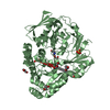
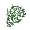
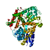
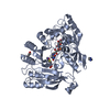
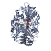
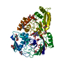
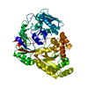
| Title | Crystal structure of Sialic acid Binding protein from Haemophilus ducreyi with Neu5Ac | |||||||||
 Components Components | Putative ABC transporter periplasmic binding protein | |||||||||
 Keywords Keywords | SUGAR BINDING PROTEIN / three domain proteins / sialic acid Neu5Gc / bacteria / molecular mimicry / adhesion / cell signaling / nutrition | |||||||||
| Function / homology |  Function and homology information Function and homology informationpeptide transport / peptide transmembrane transporter activity / ATP-binding cassette (ABC) transporter complex / outer membrane-bounded periplasmic space Similarity search - Function | |||||||||
| Biological species |  Haemophilus ducreyi 35000HP (bacteria) Haemophilus ducreyi 35000HP (bacteria) | |||||||||
| Method |  X-RAY DIFFRACTION / SYNCHROTRON / MOLECULAR REPLACEMENT / Resolution: 1.494 Å X-RAY DIFFRACTION / SYNCHROTRON / MOLECULAR REPLACEMENT / Resolution: 1.494 Å | |||||||||
 Authors Authors | Subramanian, R. / Setty, T.G. | |||||||||
| Funding support |  India, 2items India, 2items
| |||||||||
 Citation Citation | Journal: J. Biol. Chem. / Year: 2018 Title: Molecular characterization of the interaction of sialic acid with the periplasmic binding protein fromHaemophilus ducreyi. Authors: Gangi Setty, T. / Mowers, J.C. / Hobbs, A.G. / Maiya, S.P. / Syed, S. / Munson Jr., R.S. / Apicella, M.A. / Subramanian, R. | |||||||||
| History |
|
- Structure visualization
| Structure viewer | Molecule: MolmilJmol/JSmol |
|---|
- Downloads & links
-Download
| PDBx/mmCIF format | 5z99.cif.gz | 127.8 KB | Display | PDBx/mmCIF format |
|---|---|---|---|---|
| PDB format | pdb5z99.ent.gz | 96.1 KB | Display | PDB format |
| PDBx/mmJSON format | 5z99.json.gz | Tree view | PDBx/mmJSON format | |
| Others |  Other downloads Other downloads |
-Validation report
| Arichive directory | https://data.pdbj.org/pub/pdb/validation_reports/z9/5z99ftp://data.pdbj.org/pub/pdb/validation_reports/z9/5z99 | HTTPS FTP |
|---|
-Related structure data
| Related structure data | 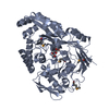 5yybSC 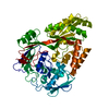 5za4C S: Starting model for refinement C: citing same article ( |
|---|---|
| Similar structure data |


-Links
 PDBj
PDBj

- Assembly
| Deposited unit | 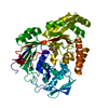
| ||||||||
|---|---|---|---|---|---|---|---|---|---|
| 1 |
| ||||||||
| Unit cell |
|
-Components
| #1: Protein | Mass: 54069.418 Da / Num. of mol.: 1 Source method: isolated from a genetically manipulated source Source: (gene. exp.) Haemophilus ducreyi 35000HP (bacteria) / Strain: 35000HP / ATCC 700724 / Gene: HD_1669 / Production host: |
|---|---|
| #2: Sugar | ChemComp-SLB /   Type: D-saccharide, beta linking / Mass: 309.270 Da / Num. of mol.: 1 Type: D-saccharide, beta linking / Mass: 309.270 Da / Num. of mol.: 1Source method: isolated from a genetically manipulated source Formula: C11H19NO9 |
| #3: Water | ChemComp-HOH /  Mass: 18.015 Da / Num. of mol.: 562 / Source method: isolated from a natural source / Formula: H2O Mass: 18.015 Da / Num. of mol.: 562 / Source method: isolated from a natural source / Formula: H2O |
-Experimental details
-Experiment
| Experiment | Method: X-RAY DIFFRACTION / Number of used crystals: 1 |
|---|
- Sample preparation
| Crystal | Density Matthews: 2.76 Å3/Da / Density % sol: 55.43 % |
|---|---|
| Crystal grow | Temperature: 291 K / Method: vapor diffusion, hanging drop / pH: 6.2 / Details: 20mM MES, 60mM NaCl, 30% PEG 1500, pH 6.2 |
-Data collection
| Diffraction | Mean temperature: 100 K | ||||||||||||||||||||||||||||||
|---|---|---|---|---|---|---|---|---|---|---|---|---|---|---|---|---|---|---|---|---|---|---|---|---|---|---|---|---|---|---|---|
| Diffraction source | Source: SYNCHROTRON / Site: APS  / Beamline: 17-ID / Wavelength: 0.9793 Å / Beamline: 17-ID / Wavelength: 0.9793 Å | ||||||||||||||||||||||||||||||
| Detector | Type: MAR CCD 130 mm / Detector: CCD / Date: Jul 7, 2006 | ||||||||||||||||||||||||||||||
| Radiation | Protocol: SINGLE WAVELENGTH / Monochromatic (M) / Laue (L): M / Scattering type: x-ray | ||||||||||||||||||||||||||||||
| Radiation wavelength | Wavelength: 0.9793 Å / Relative weight: 1 | ||||||||||||||||||||||||||||||
| Reflection | Resolution: 1.49→19.52 Å / Num. obs: 93977 / % possible obs: 96.8 % / Redundancy: 6.4 % / Biso Wilson estimate: 13.55 Å2 / CC1/2: 0.999 / Rmerge(I) obs: 0.058 / Rpim(I) all: 0.024 / Rrim(I) all: 0.063 / Net I/σ(I): 19 / Num. measured all: 604746 / Scaling rejects: 554 | ||||||||||||||||||||||||||||||
| Reflection shell | Diffraction-ID: 1
|
- Processing
| Software |
| |||||||||||||||||||||||||||||||||||||||||||||||||||||||||||||||||||||||||||||||||||||||||||||||||||||||||||||||||||||||||||||||||||||||||||||||||||||||||||||||||||||||||||||||||||||||||||||||||||||||||||||||||||||||||
|---|---|---|---|---|---|---|---|---|---|---|---|---|---|---|---|---|---|---|---|---|---|---|---|---|---|---|---|---|---|---|---|---|---|---|---|---|---|---|---|---|---|---|---|---|---|---|---|---|---|---|---|---|---|---|---|---|---|---|---|---|---|---|---|---|---|---|---|---|---|---|---|---|---|---|---|---|---|---|---|---|---|---|---|---|---|---|---|---|---|---|---|---|---|---|---|---|---|---|---|---|---|---|---|---|---|---|---|---|---|---|---|---|---|---|---|---|---|---|---|---|---|---|---|---|---|---|---|---|---|---|---|---|---|---|---|---|---|---|---|---|---|---|---|---|---|---|---|---|---|---|---|---|---|---|---|---|---|---|---|---|---|---|---|---|---|---|---|---|---|---|---|---|---|---|---|---|---|---|---|---|---|---|---|---|---|---|---|---|---|---|---|---|---|---|---|---|---|---|---|---|---|---|---|---|---|---|---|---|---|---|---|---|---|---|---|---|---|---|
| Refinement | Method to determine structure: MOLECULAR REPLACEMENT Starting model: 5YYB Resolution: 1.494→19.518 Å / SU ML: 0.15 / Cross valid method: THROUGHOUT / σ(F): 1.38 / Phase error: 18.36 / Stereochemistry target values: ML
| |||||||||||||||||||||||||||||||||||||||||||||||||||||||||||||||||||||||||||||||||||||||||||||||||||||||||||||||||||||||||||||||||||||||||||||||||||||||||||||||||||||||||||||||||||||||||||||||||||||||||||||||||||||||||
| Solvent computation | Shrinkage radii: 0.9 Å / VDW probe radii: 1.11 Å / Solvent model: FLAT BULK SOLVENT MODEL | |||||||||||||||||||||||||||||||||||||||||||||||||||||||||||||||||||||||||||||||||||||||||||||||||||||||||||||||||||||||||||||||||||||||||||||||||||||||||||||||||||||||||||||||||||||||||||||||||||||||||||||||||||||||||
| Displacement parameters | Biso max: 67.3 Å2 / Biso mean: 18.1749 Å2 / Biso min: 6.97 Å2 | |||||||||||||||||||||||||||||||||||||||||||||||||||||||||||||||||||||||||||||||||||||||||||||||||||||||||||||||||||||||||||||||||||||||||||||||||||||||||||||||||||||||||||||||||||||||||||||||||||||||||||||||||||||||||
| Refinement step | Cycle: final / Resolution: 1.494→19.518 Å
| |||||||||||||||||||||||||||||||||||||||||||||||||||||||||||||||||||||||||||||||||||||||||||||||||||||||||||||||||||||||||||||||||||||||||||||||||||||||||||||||||||||||||||||||||||||||||||||||||||||||||||||||||||||||||
| Refine LS restraints |
| |||||||||||||||||||||||||||||||||||||||||||||||||||||||||||||||||||||||||||||||||||||||||||||||||||||||||||||||||||||||||||||||||||||||||||||||||||||||||||||||||||||||||||||||||||||||||||||||||||||||||||||||||||||||||
| LS refinement shell | Refine-ID: X-RAY DIFFRACTION / Rfactor Rfree error: 0 / Total num. of bins used: 30
|
