 Movie
Movie Controller

[English] 日本語
 Yorodumi
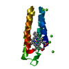
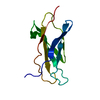
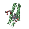
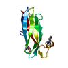
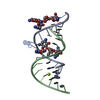
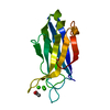
- PDB-5yo3: Crystal Structure of B562RIL with engineered disulfide bond V16C-A29C -
+ Open data

- Basic information
| Entry | Database: PDB / ID: 5yo3 | ||||||
|---|---|---|---|---|---|---|---|
| Title | Crystal Structure of B562RIL with engineered disulfide bond V16C-A29C | ||||||
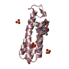
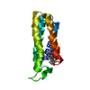
 Components Components | Soluble cytochrome b562 | ||||||
 Keywords Keywords | ELECTRON TRANSPORT / hemoprotein / fusion partner / helix bundle | ||||||
| Function / homology | Cytochrome b562 / Cytochrome b562 / Cytochrome c/b562 / electron transport chain / periplasmic space / electron transfer activity / iron ion binding / heme binding / Soluble cytochrome b562 Function and homology information Function and homology information | ||||||
| Biological species |  | ||||||
| Method |  X-RAY DIFFRACTION / SYNCHROTRON / MOLECULAR REPLACEMENT / Resolution: 1.7 Å X-RAY DIFFRACTION / SYNCHROTRON / MOLECULAR REPLACEMENT / Resolution: 1.7 Å | ||||||
 Authors Authors | Pu, M. / Xu, Z. / Song, G. / Liu, Z.J. | ||||||
 Citation Citation | Journal: Protein Cell / Year: 2018 Title: Protein crystal quality oriented disulfide bond engineering. Authors: Pu, M. / Xu, Z. / Peng, Y. / Hou, Y. / Liu, D. / Wang, Y. / Liu, H. / Song, G. / Liu, Z.J. | ||||||
| History |
|
- Structure visualization
| Structure viewer | Molecule: MolmilJmol/JSmol |
|---|
- Downloads & links
-Download
| PDBx/mmCIF format | 5yo3.cif.gz | 61 KB | Display | PDBx/mmCIF format |
|---|---|---|---|---|
| PDB format | pdb5yo3.ent.gz | 43.7 KB | Display | PDB format |
| PDBx/mmJSON format | 5yo3.json.gz | Tree view | PDBx/mmJSON format | |
| Others |  Other downloads Other downloads |
-Validation report
| Arichive directory | https://data.pdbj.org/pub/pdb/validation_reports/yo/5yo3ftp://data.pdbj.org/pub/pdb/validation_reports/yo/5yo3 | HTTPS FTP |
|---|
-Related structure data
| Related structure data | 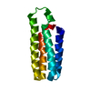 5ym7C 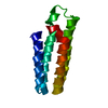 5yo4C  5yo5C 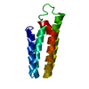 5yo6C  5yobC  5yocC  5yoeC  5yogC 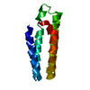 1m6tS S: Starting model for refinement C: citing same article ( |
|---|---|
| Similar structure data |

-Links
 PDBj
PDBj


- Assembly
| Deposited unit | 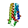
| ||||||||||||||||||
|---|---|---|---|---|---|---|---|---|---|---|---|---|---|---|---|---|---|---|---|
| 1 |
| ||||||||||||||||||
| 2 | 
| ||||||||||||||||||
| Unit cell |
| ||||||||||||||||||
| Components on special symmetry positions |
|
-Components
| #1: Protein | Mass: 11908.384 Da / Num. of mol.: 1 / Mutation: M7W, V16C, A29C, H102I, R106L Source method: isolated from a genetically manipulated source Source: (gene. exp.) | ||||
|---|---|---|---|---|---|
| #2: Chemical | ChemComp-SO4 /   Mass: 96.063 Da / Num. of mol.: 4 / Source method: obtained synthetically / Formula: SO4 Mass: 96.063 Da / Num. of mol.: 4 / Source method: obtained synthetically / Formula: SO4#3: Water | ChemComp-HOH / |  Mass: 18.015 Da / Num. of mol.: 196 / Source method: isolated from a natural source / Formula: H2O Mass: 18.015 Da / Num. of mol.: 196 / Source method: isolated from a natural source / Formula: H2OHas protein modification | Y | |
-Experimental details
-Experiment
| Experiment | Method: X-RAY DIFFRACTION / Number of used crystals: 1 |
|---|
- Sample preparation
| Crystal | Density Matthews: 2 Å3/Da / Density % sol: 38.4 % |
|---|---|
| Crystal grow | Temperature: 292 K / Method: vapor diffusion, hanging drop / pH: 9 / Details: NH4(SO4), Bicine, N-octanoylsucrose / PH range: 9.0-9.4 |
-Data collection
| Diffraction | Mean temperature: 100 K / Ambient temp details: Liquid nitrogen |
|---|---|
| Diffraction source | Source: SYNCHROTRON / Site: SSRF  / Beamline: BL17U1 / Wavelength: 0.9793 Å / Beamline: BL17U1 / Wavelength: 0.9793 Å |
| Detector | Type: MAR CCD 130 mm / Detector: CCD / Date: Jul 15, 2016 |
| Radiation | Protocol: SINGLE WAVELENGTH / Monochromatic (M) / Laue (L): M / Scattering type: x-ray |
| Radiation wavelength | Wavelength: 0.9793 Å / Relative weight: 1 |
| Reflection | Resolution: 1.7→47.118 Å / Num. obs: 10312 / % possible obs: 95 % / Redundancy: 4.4 % / Biso Wilson estimate: 16.76 Å2 / Net I/σ(I): 55.71 |
| Reflection shell | Resolution: 1.7→1.78 Å |
- Processing
| Software |
| ||||||||||||||||||||||||||||||||||||||||||||||||||||||||
|---|---|---|---|---|---|---|---|---|---|---|---|---|---|---|---|---|---|---|---|---|---|---|---|---|---|---|---|---|---|---|---|---|---|---|---|---|---|---|---|---|---|---|---|---|---|---|---|---|---|---|---|---|---|---|---|---|---|
| Refinement | Method to determine structure: MOLECULAR REPLACEMENT Starting model: 1M6T Resolution: 1.7→47.118 Å / SU ML: 0.17 / Cross valid method: FREE R-VALUE / σ(F): 1.38 / Phase error: 18.94 / Stereochemistry target values: ML
| ||||||||||||||||||||||||||||||||||||||||||||||||||||||||
| Solvent computation | Shrinkage radii: 0.9 Å / VDW probe radii: 1.11 Å / Solvent model: FLAT BULK SOLVENT MODEL | ||||||||||||||||||||||||||||||||||||||||||||||||||||||||
| Refinement step | Cycle: LAST / Resolution: 1.7→47.118 Å
| ||||||||||||||||||||||||||||||||||||||||||||||||||||||||
| Refine LS restraints |
| ||||||||||||||||||||||||||||||||||||||||||||||||||||||||
| LS refinement shell |
| ||||||||||||||||||||||||||||||||||||||||||||||||||||||||
| Refinement TLS params. | Method: refined / Origin x: 16.4812 Å / Origin y: 3.0592 Å / Origin z: -11.2233 Å
| ||||||||||||||||||||||||||||||||||||||||||||||||||||||||
| Refinement TLS group | Selection details: chain 'A' and resid 0 through 106 |
