| Entry | Database: PDB / ID: 5yo2
|
|---|
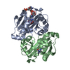
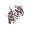
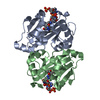
| Title | The crystal structure of Rv2747 from Mycobacterium tuberculosis in complex with Acetyl CoA and L-Arginine |
|---|
 Components Components | Amino-acid acetyltransferase |
|---|
 Keywords Keywords | TRANSFERASE / Acetyltransferase |
|---|
| Function / homology |  Function and homology information Function and homology information
amino-acid N-acetyltransferase / L-glutamate N-acetyltransferase activity, acting on acetyl-CoA as donor / : / N-acetyltransferase activity / protein homotetramerization / protein homodimerization activity / cytoplasmSimilarity search - Function Acetyltransferase NSI-like / Acetyltransferase (GNAT) family / Gcn5-related N-acetyltransferase (GNAT) domain profile. / GNAT domain / Acyl-CoA N-acyltransferaseSimilarity search - Domain/homology |
|---|
| Biological species |  Mycobacterium tuberculosis H37Rv (bacteria) Mycobacterium tuberculosis H37Rv (bacteria) |
|---|
| Method |  X-RAY DIFFRACTION / MOLECULAR REPLACEMENT / Resolution: 2.997 Å X-RAY DIFFRACTION / MOLECULAR REPLACEMENT / Resolution: 2.997 Å |
|---|
 Authors Authors | Singh, E. / Tiruttani Subhramanyam, U.K. / Pal, R.K. / Srinivasan, A. / Gourinath, S. / Das, U. |
|---|
 Citation Citation | Journal: Int. J. Biol. Macromol. / Year: 2019
Title: Structural insights into the substrate binding mechanism of novel ArgA from Mycobacterium tuberculosis
Authors: Das, U. / Singh, E. / Dharavath, S. / Tiruttani Subhramanyam, U.K. / Pal, R.K. / Vijayan, R. / Menon, S. / Kumar, S. / Gourinath, S. / Srinivasan, A. |
|---|
| History | | Deposition | Oct 26, 2017 | Deposition site: PDBJ / Processing site: PDBJ |
|---|
| Revision 1.0 | Nov 7, 2018 | Provider: repository / Type: Initial release |
|---|
| Revision 1.1 | Dec 26, 2018 | Group: Data collection / Database references / Category: citation / citation_author
Item: _citation.country / _citation.journal_abbrev ..._citation.country / _citation.journal_abbrev / _citation.journal_id_ASTM / _citation.journal_id_CSD / _citation.journal_id_ISSN / _citation.pdbx_database_id_DOI / _citation.title / _citation.year |
|---|
| Revision 1.2 | Jan 16, 2019 | Group: Data collection / Database references / Category: citation / citation_author
Item: _citation.journal_abbrev / _citation.journal_id_ISSN ..._citation.journal_abbrev / _citation.journal_id_ISSN / _citation.pdbx_database_id_PubMed / _citation_author.name |
|---|
| Revision 1.3 | Feb 13, 2019 | Group: Data collection / Database references / Category: citation / citation_author
Item: _citation.journal_volume / _citation.page_first ..._citation.journal_volume / _citation.page_first / _citation.page_last / _citation.year / _citation_author.name |
|---|
| Revision 1.4 | Mar 27, 2024 | Group: Data collection / Database references / Category: chem_comp_atom / chem_comp_bond / database_2
Item: _database_2.pdbx_DOI / _database_2.pdbx_database_accession |
|---|
|
|---|
 Movie
Movie Controller

 Yorodumi
 Open data
 Basic information
 Structure visualization
 Downloads & links
 Other downloads


 PDBj
PDBj


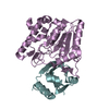
 Assembly