 Movie
Movie Controller


+ Open data

- Basic information
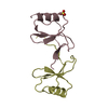
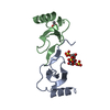
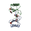
| Entry | Database: PDB / ID: 5yhr | ||||||
|---|---|---|---|---|---|---|---|
| Title | Crystal structure of the anti-CRISPR protein, AcrF2 | ||||||
 Components Components | Anti-CRISPR protein 30 | ||||||
 Keywords Keywords | VIRAL PROTEIN / anti-CRISPR protein | ||||||
| Function / homology | symbiont-mediated suppression of host CRISPR-cas system / symbiont-mediated suppression of host adaptive immune response / virus-mediated perturbation of host defense response / Anti-CRISPR protein 30 Function and homology information Function and homology information | ||||||
| Biological species |  Pseudomonas phage D3112 (virus) Pseudomonas phage D3112 (virus) | ||||||
| Method |  X-RAY DIFFRACTION / SYNCHROTRON / SAD / Resolution: 1.34 Å X-RAY DIFFRACTION / SYNCHROTRON / SAD / Resolution: 1.34 Å | ||||||
 Authors Authors | Hong, S. / Ka, D. / Bae, E. | ||||||
 Citation Citation | Journal: J. Biol. Chem. / Year: 2018 Title: CRISPR RNA and anti-CRISPR protein binding to theXanthomonas albilineansCsy1-Csy2 heterodimer in the type I-F CRISPR-Cas system Authors: Hong, S. / Ka, D. / Yoon, S.J. / Suh, N. / Jeong, M. / Suh, J.Y. / Bae, E. | ||||||
| History |
|
- Structure visualization
| Structure viewer | Molecule: MolmilJmol/JSmol |
|---|
- Downloads & links
-Download
| PDBx/mmCIF format | 5yhr.cif.gz | 86.2 KB | Display | PDBx/mmCIF format |
|---|---|---|---|---|
| PDB format | pdb5yhr.ent.gz | 68.8 KB | Display | PDB format |
| PDBx/mmJSON format | 5yhr.json.gz | Tree view | PDBx/mmJSON format | |
| Others |  Other downloads Other downloads |
-Validation report
| Summary document | 5yhr_validation.pdf.gz | 422.3 KB | Display | wwPDB validaton report |
|---|---|---|---|---|
| Full document | 5yhr_full_validation.pdf.gz | 422.7 KB | Display | |
| Data in XML | 5yhr_validation.xml.gz | 11.9 KB | Display | |
| Data in CIF | 5yhr_validation.cif.gz | 17.6 KB | Display | |
| Arichive directory | https://data.pdbj.org/pub/pdb/validation_reports/yh/5yhrftp://data.pdbj.org/pub/pdb/validation_reports/yh/5yhr | HTTPS FTP |
-Related structure data
| Similar structure data |
|---|


-Links
 PDBj
PDBj- Assembly
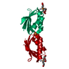
| Deposited unit | 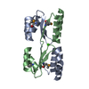
| ||||||||
|---|---|---|---|---|---|---|---|---|---|
| 1 | 
| ||||||||
| 2 | 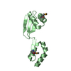
| ||||||||
| 3 |
| ||||||||
| Unit cell |
|
-Components
| #1: Protein | Mass: 10791.127 Da / Num. of mol.: 2 Source method: isolated from a genetically manipulated source Source: (gene. exp.) Pseudomonas phage D3112 (virus) / Gene: orf30 / Production host:  #2: Chemical | ChemComp-CA / |   Mass: 40.078 Da / Num. of mol.: 1 / Source method: obtained synthetically / Formula: Ca Mass: 40.078 Da / Num. of mol.: 1 / Source method: obtained synthetically / Formula: Ca#3: Water | ChemComp-HOH / |  Mass: 18.015 Da / Num. of mol.: 302 / Source method: isolated from a natural source / Formula: H2O Mass: 18.015 Da / Num. of mol.: 302 / Source method: isolated from a natural source / Formula: H2O |
|---|
-Experimental details
-Experiment
| Experiment | Method: X-RAY DIFFRACTION / Number of used crystals: 1 |
|---|
- Sample preparation
| Crystal | Density Matthews: 2.55 Å3/Da / Density % sol: 51.85 % |
|---|---|
| Crystal grow | Temperature: 293 K / Method: evaporation / Details: 21% (w/v) PEG 6000, 0.4 M CaCl2, 100 mM MES pH 5.5 |
-Data collection
| Diffraction | Mean temperature: 100 K |
|---|---|
| Diffraction source | Source: SYNCHROTRON / Site: PAL/PLS  / Beamline: 7A (6B, 6C1) / Wavelength: 0.97929 Å / Beamline: 7A (6B, 6C1) / Wavelength: 0.97929 Å |
| Detector | Type: ADSC QUANTUM 270 / Detector: CCD / Date: Sep 13, 2017 |
| Radiation | Protocol: SINGLE WAVELENGTH / Monochromatic (M) / Laue (L): M / Scattering type: x-ray |
| Radiation wavelength | Wavelength: 0.97929 Å / Relative weight: 1 |
| Reflection | Resolution: 1.34→50 Å / Num. obs: 50291 / % possible obs: 100 % / Redundancy: 27.9 % / Rmerge(I) obs: 0.102 / Rpim(I) all: 0.02 / Rrim(I) all: 0.104 / Net I/σ(I): 29.79 |
| Reflection shell | Resolution: 1.34→1.39 Å / Redundancy: 25.6 % / Rmerge(I) obs: 0.582 / Mean I/σ(I) obs: 6.43 / Num. unique obs: 4925 / Rpim(I) all: 0.117 / Rrim(I) all: 0.594 / % possible all: 100 |
- Processing
| Software |
| |||||||||||||||||||||||||||||||||||||||||||||||||||||||||||||||||||||||||||||||||||||||||||||||||||||||||
|---|---|---|---|---|---|---|---|---|---|---|---|---|---|---|---|---|---|---|---|---|---|---|---|---|---|---|---|---|---|---|---|---|---|---|---|---|---|---|---|---|---|---|---|---|---|---|---|---|---|---|---|---|---|---|---|---|---|---|---|---|---|---|---|---|---|---|---|---|---|---|---|---|---|---|---|---|---|---|---|---|---|---|---|---|---|---|---|---|---|---|---|---|---|---|---|---|---|---|---|---|---|---|---|---|---|---|
| Refinement | Method to determine structure: SAD / Resolution: 1.34→23.174 Å / SU ML: 0.09 / Cross valid method: FREE R-VALUE / σ(F): 0 / Phase error: 16.13
| |||||||||||||||||||||||||||||||||||||||||||||||||||||||||||||||||||||||||||||||||||||||||||||||||||||||||
| Solvent computation | Shrinkage radii: 0.9 Å / VDW probe radii: 1.11 Å | |||||||||||||||||||||||||||||||||||||||||||||||||||||||||||||||||||||||||||||||||||||||||||||||||||||||||
| Refinement step | Cycle: LAST / Resolution: 1.34→23.174 Å
| |||||||||||||||||||||||||||||||||||||||||||||||||||||||||||||||||||||||||||||||||||||||||||||||||||||||||
| Refine LS restraints |
| |||||||||||||||||||||||||||||||||||||||||||||||||||||||||||||||||||||||||||||||||||||||||||||||||||||||||
| LS refinement shell |
|
