 Movie
Movie Controller

[English] 日本語
 Yorodumi
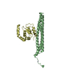
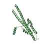
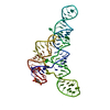
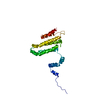
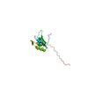
- PDB-5y9r: Crystal structure of the oligomerization domain of NSP4 from rota... -
+ Open data

- Basic information
| Entry | Database: PDB / ID: 5y9r | ||||||
|---|---|---|---|---|---|---|---|
| Title | Crystal structure of the oligomerization domain of NSP4 from rotavirus strain MF66 | ||||||
 Components Components | Nonstructural protein 4 | ||||||
 Keywords Keywords | VIRAL PROTEIN / antiparallel / tetramer / coiled-coil / nickel bound | ||||||
| Function / homology |  Function and homology information Function and homology informationhost caveola / host cell rough endoplasmic reticulum membrane / viral process / channel activity / toxin activity / monoatomic ion transmembrane transport / symbiont-mediated activation of host autophagy / extracellular region / metal ion binding / membrane Similarity search - Function | ||||||
| Biological species |  Bovine rotavirus G10 Bovine rotavirus G10 | ||||||
| Method |  X-RAY DIFFRACTION / SYNCHROTRON / MOLECULAR REPLACEMENT / Resolution: 2.2 Å X-RAY DIFFRACTION / SYNCHROTRON / MOLECULAR REPLACEMENT / Resolution: 2.2 Å | ||||||
 Authors Authors | Suguna, K. / Kumar, S. | ||||||
| Funding support |  India, 1items India, 1items
| ||||||
 Citation Citation | Journal: To Be Published Title: New tetrameric forms of the rotavirus NSP4 with antiparallel helices Authors: Suguna, K. / Kumar, S. | ||||||
| History |
|
- Structure visualization
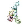
| Structure viewer | Molecule: MolmilJmol/JSmol |
|---|
- Downloads & links
-Download
| PDBx/mmCIF format | 5y9r.cif.gz | 47.1 KB | Display | PDBx/mmCIF format |
|---|---|---|---|---|
| PDB format | pdb5y9r.ent.gz | 32.9 KB | Display | PDB format |
| PDBx/mmJSON format | 5y9r.json.gz | Tree view | PDBx/mmJSON format | |
| Others |  Other downloads Other downloads |
-Validation report
| Summary document | 5y9r_validation.pdf.gz | 458.4 KB | Display | wwPDB validaton report |
|---|---|---|---|---|
| Full document | 5y9r_full_validation.pdf.gz | 458.9 KB | Display | |
| Data in XML | 5y9r_validation.xml.gz | 8.4 KB | Display | |
| Data in CIF | 5y9r_validation.cif.gz | 10.7 KB | Display | |
| Arichive directory | https://data.pdbj.org/pub/pdb/validation_reports/y9/5y9rftp://data.pdbj.org/pub/pdb/validation_reports/y9/5y9r | HTTPS FTP |
-Related structure data
| Similar structure data |
|---|
-Links
 PDBj
PDBj- Assembly
| Deposited unit | 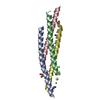
| ||||||||
|---|---|---|---|---|---|---|---|---|---|
| 1 | 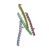
| ||||||||
| Unit cell |
|
-Components
| #1: Protein/peptide | Mass: 5649.737 Da / Num. of mol.: 4 / Fragment: UNP residues 95-139 Source method: isolated from a genetically manipulated source Source: (gene. exp.) Bovine rotavirus G10 / Production host:  #2: Chemical |   Mass: 58.693 Da / Num. of mol.: 2 / Source method: obtained synthetically / Formula: Ni Mass: 58.693 Da / Num. of mol.: 2 / Source method: obtained synthetically / Formula: Ni#3: Chemical | ChemComp-EDO /   Mass: 62.068 Da / Num. of mol.: 4 / Source method: obtained synthetically / Formula: C2H6O2 Mass: 62.068 Da / Num. of mol.: 4 / Source method: obtained synthetically / Formula: C2H6O2#4: Chemical | ChemComp-CD / |   Mass: 112.411 Da / Num. of mol.: 1 / Source method: obtained synthetically / Formula: Cd Mass: 112.411 Da / Num. of mol.: 1 / Source method: obtained synthetically / Formula: Cd#5: Water | ChemComp-HOH / |  Mass: 18.015 Da / Num. of mol.: 41 / Source method: isolated from a natural source / Formula: H2O Mass: 18.015 Da / Num. of mol.: 41 / Source method: isolated from a natural source / Formula: H2O |
|---|
-Experimental details
-Experiment
| Experiment | Method: X-RAY DIFFRACTION / Number of used crystals: 1 |
|---|
- Sample preparation
| Crystal | Density Matthews: 2.03 Å3/Da / Density % sol: 39.5 % |
|---|---|
| Crystal grow | Temperature: 299 K / Method: microbatch / pH: 4.5 Details: 0.1M sodium acetate trihydrate, 24% PEG 2000, 10mM magnesium chloride, 10mM nickel chloride, 10mM cadmium chloride |
-Data collection
| Diffraction | Mean temperature: 100 K |
|---|---|
| Diffraction source | Source: SYNCHROTRON / Site: ESRF  / Beamline: BM14 / Wavelength: 0.9537 Å / Beamline: BM14 / Wavelength: 0.9537 Å |
| Detector | Type: MARMOSAIC 225 mm CCD / Detector: CCD / Date: Apr 29, 2015 / Details: bent collimating mirror and toroid |
| Radiation | Protocol: SINGLE WAVELENGTH / Monochromatic (M) / Laue (L): M / Scattering type: x-ray |
| Radiation wavelength | Wavelength: 0.9537 Å / Relative weight: 1 |
| Reflection | Resolution: 2.2→62.6 Å / Num. obs: 9675 / % possible obs: 98.8 % / Redundancy: 4.9 % / CC1/2: 0.997 / Rmerge(I) obs: 0.063 / Rpim(I) all: 0.047 / Rrim(I) all: 0.079 / Net I/σ(I): 12.8 |
| Reflection shell | Resolution: 2.2→2.27 Å / Redundancy: 4.9 % / Rmerge(I) obs: 0.41 / Mean I/σ(I) obs: 3.5 / Num. unique obs: 821 / CC1/2: 0.875 / Rpim(I) all: 0.31 / Rrim(I) all: 0.52 / % possible all: 99.3 |
- Processing
| Software |
| |||||||||||||||||||||||||||||||||||||||||||||||||
|---|---|---|---|---|---|---|---|---|---|---|---|---|---|---|---|---|---|---|---|---|---|---|---|---|---|---|---|---|---|---|---|---|---|---|---|---|---|---|---|---|---|---|---|---|---|---|---|---|---|---|
| Refinement | Method to determine structure: MOLECULAR REPLACEMENT / Resolution: 2.2→39.595 Å / SU ML: 0.22 / Cross valid method: FREE R-VALUE / σ(F): 1.34 / Phase error: 26.06
| |||||||||||||||||||||||||||||||||||||||||||||||||
| Solvent computation | Shrinkage radii: 0.9 Å / VDW probe radii: 1.11 Å | |||||||||||||||||||||||||||||||||||||||||||||||||
| Refinement step | Cycle: LAST / Resolution: 2.2→39.595 Å
| |||||||||||||||||||||||||||||||||||||||||||||||||
| Refine LS restraints |
| |||||||||||||||||||||||||||||||||||||||||||||||||
| LS refinement shell |
|
