 Movie
Movie Controller


+ Open data

- Basic information
| Entry | Database: PDB / ID: 5y38 | ||||||
|---|---|---|---|---|---|---|---|
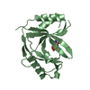
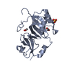
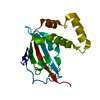
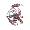
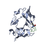
| Title | Crystal structure of C7orf59-HBXIP complex | ||||||
 Components Components |
| ||||||
 Keywords Keywords | SIGNALING PROTEIN / Ragulator / LAMTOR / HBXIP / C7orf59 | ||||||
| Function / homology |  Function and homology information Function and homology informationFNIP-folliculin RagC/D GAP / Ragulator complex / protein localization to lysosome / MTOR signalling / Energy dependent regulation of mTOR by LKB1-AMPK / Amino acids regulate mTORC1 / regulation of cell size / Macroautophagy / TORC1 signaling / mTORC1-mediated signalling ...FNIP-folliculin RagC/D GAP / Ragulator complex / protein localization to lysosome / MTOR signalling / Energy dependent regulation of mTOR by LKB1-AMPK / Amino acids regulate mTORC1 / regulation of cell size / Macroautophagy / TORC1 signaling / mTORC1-mediated signalling / positive regulation of TOR signaling / positive regulation of TORC1 signaling / viral genome replication / Regulation of PTEN gene transcription / positive regulation of interleukin-8 production / TP53 Regulates Metabolic Genes / cellular response to amino acid stimulus / positive regulation of protein localization to nucleus / response to virus / late endosome membrane / positive regulation of canonical NF-kappaB signal transduction / lysosome / lysosomal membrane / positive regulation of gene expression / negative regulation of apoptotic process / protein-containing complex / cytosol Similarity search - Function | ||||||
| Biological species |  Homo sapiens (human) Homo sapiens (human) | ||||||
| Method |  X-RAY DIFFRACTION / SYNCHROTRON / MOLECULAR REPLACEMENT / molecular replacement / Resolution: 2.8 Å X-RAY DIFFRACTION / SYNCHROTRON / MOLECULAR REPLACEMENT / molecular replacement / Resolution: 2.8 Å | ||||||
 Authors Authors | Zhang, T. / Ding, J. | ||||||
 Citation Citation | Journal: Nat Commun / Year: 2017 Title: Structural basis for Ragulator functioning as a scaffold in membrane-anchoring of Rag GTPases and mTORC1 Authors: Zhang, T. / Wang, R. / Wang, Z. / Wang, X. / Wang, F. / Ding, J. | ||||||
| History |
|
- Structure visualization
| Structure viewer | Molecule: MolmilJmol/JSmol |
|---|
- Downloads & links
-Download
| PDBx/mmCIF format | 5y38.cif.gz | 77.5 KB | Display | PDBx/mmCIF format |
|---|---|---|---|---|
| PDB format | pdb5y38.ent.gz | 56.6 KB | Display | PDB format |
| PDBx/mmJSON format | 5y38.json.gz | Tree view | PDBx/mmJSON format | |
| Others |  Other downloads Other downloads |
-Validation report
| Arichive directory | https://data.pdbj.org/pub/pdb/validation_reports/y3/5y38ftp://data.pdbj.org/pub/pdb/validation_reports/y3/5y38 | HTTPS FTP |
|---|
-Related structure data
| Related structure data |  5y39C  5y3aC  3ms6S S: Starting model for refinement C: citing same article ( |
|---|---|
| Similar structure data |


-Links
 PDBj
PDBj

- Assembly
| Deposited unit | 
| ||||||||
|---|---|---|---|---|---|---|---|---|---|
| 1 |
| ||||||||
| Unit cell |
|
-Components
| #1: Protein | Mass: 9622.900 Da / Num. of mol.: 1 / Fragment: Roadblock domain Source method: isolated from a genetically manipulated source Source: (gene. exp.) Homo sapiens (human) / Gene: LAMTOR5, HBXIP, XIP / Plasmid: pET-Duet / Production host:  | ||
|---|---|---|---|
| #2: Protein | Mass: 12012.636 Da / Num. of mol.: 1 / Fragment: Roadblock domain Source method: isolated from a genetically manipulated source Source: (gene. exp.) Homo sapiens (human) / Gene: LAMTOR4, C7orf59 / Plasmid: pET28a / Production host: | ||
| #3: Chemical |   Mass: 96.063 Da / Num. of mol.: 3 / Source method: obtained synthetically / Formula: SO4 Mass: 96.063 Da / Num. of mol.: 3 / Source method: obtained synthetically / Formula: SO4#4: Water | ChemComp-HOH / |  Mass: 18.015 Da / Num. of mol.: 10 / Source method: isolated from a natural source / Formula: H2O Mass: 18.015 Da / Num. of mol.: 10 / Source method: isolated from a natural source / Formula: H2O |
-Experimental details
-Experiment
| Experiment | Method: X-RAY DIFFRACTION / Number of used crystals: 1 |
|---|
- Sample preparation
| Crystal | Density Matthews: 2.06 Å3/Da / Density % sol: 40.23 % |
|---|---|
| Crystal grow | Temperature: 289 K / Method: vapor diffusion, hanging drop / pH: 4.6 Details: 0.16 M ammonium sulfate, 0.08 M sodium acetate trihydrate (pH 4.6), and 20% (w/v) PEG 4000 |
-Data collection
| Diffraction | Mean temperature: 100 K | ||||||||||||||||||||||||||||||||||||||||||||||||||||||||||||||||||
|---|---|---|---|---|---|---|---|---|---|---|---|---|---|---|---|---|---|---|---|---|---|---|---|---|---|---|---|---|---|---|---|---|---|---|---|---|---|---|---|---|---|---|---|---|---|---|---|---|---|---|---|---|---|---|---|---|---|---|---|---|---|---|---|---|---|---|---|
| Diffraction source | Source: SYNCHROTRON / Site: SSRF  / Beamline: BL17U1 / Wavelength: 1 Å / Beamline: BL17U1 / Wavelength: 1 Å | ||||||||||||||||||||||||||||||||||||||||||||||||||||||||||||||||||
| Detector | Type: RAYONIX MX225HE / Detector: CCD / Date: May 6, 2015 | ||||||||||||||||||||||||||||||||||||||||||||||||||||||||||||||||||
| Radiation | Protocol: SINGLE WAVELENGTH / Monochromatic (M) / Laue (L): M / Scattering type: x-ray | ||||||||||||||||||||||||||||||||||||||||||||||||||||||||||||||||||
| Radiation wavelength | Wavelength: 1 Å / Relative weight: 1 | ||||||||||||||||||||||||||||||||||||||||||||||||||||||||||||||||||
| Reflection | Resolution: 2.8→50 Å / Num. obs: 4579 / % possible obs: 97.1 % / Redundancy: 6.3 % / Rmerge(I) obs: 0.095 / Χ2: 1.403 / Net I/σ(I): 9.3 | ||||||||||||||||||||||||||||||||||||||||||||||||||||||||||||||||||
| Reflection shell |
|
-Phasing
| Phasing | Method: molecular replacement |
|---|
- Processing
| Software |
| |||||||||||||||||||||||||||||||||||||||||||||||||||||||||||||||||||||||||||
|---|---|---|---|---|---|---|---|---|---|---|---|---|---|---|---|---|---|---|---|---|---|---|---|---|---|---|---|---|---|---|---|---|---|---|---|---|---|---|---|---|---|---|---|---|---|---|---|---|---|---|---|---|---|---|---|---|---|---|---|---|---|---|---|---|---|---|---|---|---|---|---|---|---|---|---|---|
| Refinement | Method to determine structure: MOLECULAR REPLACEMENT Starting model: 3MS6 Resolution: 2.8→50 Å / Cor.coef. Fo:Fc: 0.929 / Cor.coef. Fo:Fc free: 0.92 / SU B: 32.485 / SU ML: 0.277 / Cross valid method: THROUGHOUT / σ(F): 0 / ESU R Free: 0.39 / Details: U VALUES : WITH TLS ADDED
| |||||||||||||||||||||||||||||||||||||||||||||||||||||||||||||||||||||||||||
| Solvent computation | Ion probe radii: 0.8 Å / Shrinkage radii: 0.8 Å / VDW probe radii: 1.2 Å | |||||||||||||||||||||||||||||||||||||||||||||||||||||||||||||||||||||||||||
| Displacement parameters | Biso max: 133.51 Å2 / Biso mean: 50.674 Å2 / Biso min: 25.05 Å2
| |||||||||||||||||||||||||||||||||||||||||||||||||||||||||||||||||||||||||||
| Refinement step | Cycle: final / Resolution: 2.8→50 Å
| |||||||||||||||||||||||||||||||||||||||||||||||||||||||||||||||||||||||||||
| Refine LS restraints |
| |||||||||||||||||||||||||||||||||||||||||||||||||||||||||||||||||||||||||||
| LS refinement shell | Resolution: 2.8→2.873 Å / Rfactor Rfree error: 0 / Total num. of bins used: 20
| |||||||||||||||||||||||||||||||||||||||||||||||||||||||||||||||||||||||||||
| Refinement TLS params. | Method: refined / Refine-ID: X-RAY DIFFRACTION
| |||||||||||||||||||||||||||||||||||||||||||||||||||||||||||||||||||||||||||
| Refinement TLS group |
|
