 Movie
Movie Controller

[English] 日本語
 Yorodumi
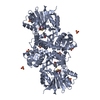
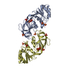
- PDB-5xvs: Crystal structure of UDP-GlcNAc 2-epimerase NeuC complexed with UDP -
+ Open data

- Basic information
| Entry | Database: PDB / ID: 5xvs | ||||||
|---|---|---|---|---|---|---|---|
| Title | Crystal structure of UDP-GlcNAc 2-epimerase NeuC complexed with UDP | ||||||
 Components Components | GDP/UDP-N,N'-diacetylbacillosamine 2-epimerase (Hydrolyzing) | ||||||
 Keywords Keywords | HYDROLASE / NeuC / UDP N-acetylglucosamine 2-epimerase / TRANSFERASE | ||||||
| Function / homology |  Function and homology information Function and homology informationUDP-N,N'-diacetylbacillosamine 2-epimerase (hydrolysing) / UDP-N,N'-diacetylbacillosamine 2-epimerase activity / UDP-N-acetylglucosamine 2-epimerase (hydrolysing) / UDP-N-acetylglucosamine metabolic process / nucleotide binding Similarity search - Function | ||||||
| Biological species |  Acinetobacter baumannii (bacteria) Acinetobacter baumannii (bacteria) | ||||||
| Method |  X-RAY DIFFRACTION / SYNCHROTRON / MOLECULAR REPLACEMENT / Resolution: 2.383 Å X-RAY DIFFRACTION / SYNCHROTRON / MOLECULAR REPLACEMENT / Resolution: 2.383 Å | ||||||
 Authors Authors | Ko, T.P. / Hsieh, T.J. / Chen, S.C. / Wu, S.C. / Guan, H.H. / Yang, C.H. / Chen, C.J. / Chen, Y. | ||||||
 Citation Citation | Journal: J. Biol. Chem. / Year: 2018 Title: The tetrameric structure of sialic acid-synthesizing UDP-GlcNAc 2-epimerase fromAcinetobacter baumannii: A comparative study with human GNE. Authors: Ko, T.P. / Lai, S.J. / Hsieh, T.J. / Yang, C.S. / Chen, Y. | ||||||
| History |
|
- Structure visualization
| Structure viewer | Molecule: MolmilJmol/JSmol |
|---|
- Downloads & links
-Download
| PDBx/mmCIF format | 5xvs.cif.gz | 320.9 KB | Display | PDBx/mmCIF format |
|---|---|---|---|---|
| PDB format | pdb5xvs.ent.gz | 259.7 KB | Display | PDB format |
| PDBx/mmJSON format | 5xvs.json.gz | Tree view | PDBx/mmJSON format | |
| Others |  Other downloads Other downloads |
-Validation report
| Arichive directory | https://data.pdbj.org/pub/pdb/validation_reports/xv/5xvsftp://data.pdbj.org/pub/pdb/validation_reports/xv/5xvs | HTTPS FTP |
|---|
-Related structure data
| Related structure data |  5zlrC  5zltC  4zhtS  5h50 5h6p S: Starting model for refinement C: citing same article ( |
|---|---|
| Similar structure data |


-Links
 PDBj
PDBj


- Assembly
| Deposited unit | 
| |||||||||||||||||||||||||||
|---|---|---|---|---|---|---|---|---|---|---|---|---|---|---|---|---|---|---|---|---|---|---|---|---|---|---|---|---|
| 1 |
| |||||||||||||||||||||||||||
| Unit cell |
| |||||||||||||||||||||||||||
| Components on special symmetry positions |
|
-Components
| #1: Protein | Mass: 42300.414 Da / Num. of mol.: 2 Source method: isolated from a genetically manipulated source Source: (gene. exp.) Acinetobacter baumannii (bacteria) / Gene: legG, LV38_02406 / Production host: References: UniProt: A0A154EJU5, UDP-N,N'-diacetylbacillosamine 2-epimerase (hydrolysing) #2: Chemical |   Mass: 6.941 Da / Num. of mol.: 2 / Source method: isolated from a natural source / Formula: Li Mass: 6.941 Da / Num. of mol.: 2 / Source method: isolated from a natural source / Formula: Li#3: Chemical |   Type: RNA linking / Mass: 404.161 Da / Num. of mol.: 2 / Source method: obtained synthetically / Formula: C9H14N2O12P2 / Comment: UDP*YM Type: RNA linking / Mass: 404.161 Da / Num. of mol.: 2 / Source method: obtained synthetically / Formula: C9H14N2O12P2 / Comment: UDP*YM#4: Sugar | ChemComp-NAG / |   Type: D-saccharide, beta linking / Mass: 221.208 Da / Num. of mol.: 1 Type: D-saccharide, beta linking / Mass: 221.208 Da / Num. of mol.: 1Source method: isolated from a genetically manipulated source Formula: C8H15NO6 #5: Water | ChemComp-HOH / |  Mass: 18.015 Da / Num. of mol.: 423 / Source method: isolated from a natural source / Formula: H2O Mass: 18.015 Da / Num. of mol.: 423 / Source method: isolated from a natural source / Formula: H2O |
|---|
-Experimental details
-Experiment
| Experiment | Method: X-RAY DIFFRACTION / Number of used crystals: 1 |
|---|
- Sample preparation
| Crystal | Density Matthews: 2.39 Å3/Da / Density % sol: 48.46 % |
|---|---|
| Crystal grow | Temperature: 277 K / Method: vapor diffusion, hanging drop / pH: 8.5 / Details: 0.2 M Li2SO4, 0.1 M Tris-HCl pH 8.5, 25% PEG 3350 |
-Data collection
| Diffraction | Mean temperature: 100 K |
|---|---|
| Diffraction source | Source: SYNCHROTRON / Site: NSRRC  / Beamline: BL13B1 / Wavelength: 1 Å / Beamline: BL13B1 / Wavelength: 1 Å |
| Detector | Type: ADSC QUANTUM 315r / Detector: CCD / Date: Jul 12, 2016 |
| Radiation | Protocol: SINGLE WAVELENGTH / Monochromatic (M) / Laue (L): M / Scattering type: x-ray |
| Radiation wavelength | Wavelength: 1 Å / Relative weight: 1 |
| Reflection | Resolution: 2.38→27.95 Å / Num. obs: 32255 / % possible obs: 99.49 % / Redundancy: 6.5 % / Net I/σ(I): 9.22 |
- Processing
| Software |
| |||||||||||||||||||||||||||||||||||||||||||||||||||||||||||||||||||||||||||||||||||||||||||
|---|---|---|---|---|---|---|---|---|---|---|---|---|---|---|---|---|---|---|---|---|---|---|---|---|---|---|---|---|---|---|---|---|---|---|---|---|---|---|---|---|---|---|---|---|---|---|---|---|---|---|---|---|---|---|---|---|---|---|---|---|---|---|---|---|---|---|---|---|---|---|---|---|---|---|---|---|---|---|---|---|---|---|---|---|---|---|---|---|---|---|---|---|
| Refinement | Method to determine structure: MOLECULAR REPLACEMENT Starting model: 4ZHT Resolution: 2.383→27.96 Å / SU ML: 0.25 / Cross valid method: FREE R-VALUE / σ(F): 1.34 / Phase error: 22.59 / Stereochemistry target values: ML
| |||||||||||||||||||||||||||||||||||||||||||||||||||||||||||||||||||||||||||||||||||||||||||
| Solvent computation | Shrinkage radii: 0.9 Å / VDW probe radii: 1.11 Å / Solvent model: FLAT BULK SOLVENT MODEL | |||||||||||||||||||||||||||||||||||||||||||||||||||||||||||||||||||||||||||||||||||||||||||
| Refinement step | Cycle: LAST / Resolution: 2.383→27.96 Å
| |||||||||||||||||||||||||||||||||||||||||||||||||||||||||||||||||||||||||||||||||||||||||||
| Refine LS restraints |
| |||||||||||||||||||||||||||||||||||||||||||||||||||||||||||||||||||||||||||||||||||||||||||
| LS refinement shell |
| |||||||||||||||||||||||||||||||||||||||||||||||||||||||||||||||||||||||||||||||||||||||||||
| Refinement TLS params. | Method: refined / Origin x: 16.7123 Å / Origin y: 13.2566 Å / Origin z: 48.0755 Å
| |||||||||||||||||||||||||||||||||||||||||||||||||||||||||||||||||||||||||||||||||||||||||||
| Refinement TLS group | Selection details: all |
