 Movie
Movie Controller


+ Open data

- Basic information
| Entry | Database: PDB / ID: 5xv0 | ||||||
|---|---|---|---|---|---|---|---|
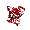
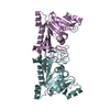
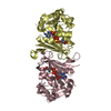
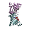
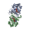
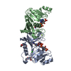
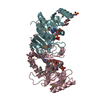
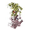
| Title | Crystal structure of Rib7 mutant D33N from Methanosarcina mazei | ||||||
 Components Components | Conserved protein | ||||||
 Keywords Keywords | OXIDOREDUCTASE / Rib7 / riboflavin biosynthesis | ||||||
| Function / homology |  Function and homology information Function and homology information2,5-diamino-6-(ribosylamino)-4(3H)-pyrimidinone 5'-phosphate reductase / 5-amino-6-(5-phosphoribosylamino)uracil reductase activity / riboflavin biosynthetic process / NADP binding Similarity search - Function | ||||||
| Biological species |  Methanosarcina mazei (archaea) Methanosarcina mazei (archaea) | ||||||
| Method |  X-RAY DIFFRACTION / SYNCHROTRON / MOLECULAR REPLACEMENT / Resolution: 1.95 Å X-RAY DIFFRACTION / SYNCHROTRON / MOLECULAR REPLACEMENT / Resolution: 1.95 Å | ||||||
 Authors Authors | Yeh, T.M. / Chen, S.C. / Chang, T.H. / Huang, M.F. / Liaw, S.H. | ||||||
 Citation Citation | Journal: Biochem. Biophys. Res. Commun. / Year: 2018 Title: Evolution of archaeal Rib7 and eubacterial RibG reductases in riboflavin biosynthesis: Substrate specificity and cofactor preference. Authors: Chen, S.C. / Yen, T.M. / Chang, T.H. / Liaw, S.H. | ||||||
| History |
|
- Structure visualization
| Structure viewer | Molecule: MolmilJmol/JSmol |
|---|
- Downloads & links
-Download
| PDBx/mmCIF format | 5xv0.cif.gz | 288.9 KB | Display | PDBx/mmCIF format |
|---|---|---|---|---|
| PDB format | pdb5xv0.ent.gz | 234.8 KB | Display | PDB format |
| PDBx/mmJSON format | 5xv0.json.gz | Tree view | PDBx/mmJSON format | |
| Others |  Other downloads Other downloads |
-Validation report
| Arichive directory | https://data.pdbj.org/pub/pdb/validation_reports/xv/5xv0ftp://data.pdbj.org/pub/pdb/validation_reports/xv/5xv0 | HTTPS FTP |
|---|
-Related structure data
| Related structure data |  5xuxSC  5xv2C 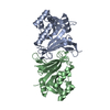 5xv5C S: Starting model for refinement C: citing same article ( |
|---|---|
| Similar structure data |


-Links
 PDBj
PDBj
- Assembly
| Deposited unit | 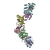
| ||||||||
|---|---|---|---|---|---|---|---|---|---|
| 1 | 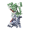
| ||||||||
| 2 | 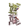
| ||||||||
| 3 | 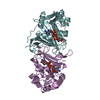
| ||||||||
| Unit cell |
| ||||||||
| Components on special symmetry positions |
|
-Components
| #1: Protein | Mass: 26104.125 Da / Num. of mol.: 6 / Mutation: D33N Source method: isolated from a genetically manipulated source Source: (gene. exp.) Methanosarcina mazei (strain ATCC BAA-159 / DSM 3647 / Goe1 / Go1 / JCM 11833 / OCM 88) (archaea)Strain: ATCC BAA-159 / DSM 3647 / Goe1 / Go1 / JCM 11833 / OCM 88 Gene: MM_0826 / Production host:  #2: Chemical | ChemComp-NAP /   Mass: 743.405 Da / Num. of mol.: 6 / Source method: obtained synthetically / Formula: C21H28N7O17P3 Mass: 743.405 Da / Num. of mol.: 6 / Source method: obtained synthetically / Formula: C21H28N7O17P3#3: Water | ChemComp-HOH / |  Mass: 18.015 Da / Num. of mol.: 699 / Source method: isolated from a natural source / Formula: H2O Mass: 18.015 Da / Num. of mol.: 699 / Source method: isolated from a natural source / Formula: H2O |
|---|
-Experimental details
-Experiment
| Experiment | Method: X-RAY DIFFRACTION / Number of used crystals: 1 |
|---|
- Sample preparation
| Crystal | Density Matthews: 3.2 Å3/Da / Density % sol: 61.62 % |
|---|---|
| Crystal grow | Temperature: 293 K / Method: vapor diffusion, hanging drop / pH: 7 Details: 10 % Tacsimate (pH 7.0), 10 % PEG 5000, and 100 mM HEPES (pH 7.0) |
-Data collection
| Diffraction | Mean temperature: 110 K |
|---|---|
| Diffraction source | Source: SYNCHROTRON / Site: NSRRC  / Beamline: BL13B1 / Wavelength: 1 Å / Beamline: BL13B1 / Wavelength: 1 Å |
| Detector | Type: ADSC QUANTUM 315r / Detector: CCD / Date: Apr 28, 2009 |
| Radiation | Protocol: SINGLE WAVELENGTH / Monochromatic (M) / Laue (L): M / Scattering type: x-ray |
| Radiation wavelength | Wavelength: 1 Å / Relative weight: 1 |
| Reflection | Resolution: 1.95→30 Å / Num. obs: 141352 / % possible obs: 99.9 % / Redundancy: 5.8 % / Net I/σ(I): 22.1 |
| Reflection shell | Resolution: 1.95→2.02 Å / Redundancy: 5.8 % / Rmerge(I) obs: 0.652 / % possible all: 100 |
- Processing
| Software |
| ||||||||||||||||||||||||||||||||||||||||||||||||||||||||||||||||||||||||||||||||||||||||||||||||||||||||||||||||||||||||||||||||||||||||||||||||||||||||||||||||||||||||||||||||||||||
|---|---|---|---|---|---|---|---|---|---|---|---|---|---|---|---|---|---|---|---|---|---|---|---|---|---|---|---|---|---|---|---|---|---|---|---|---|---|---|---|---|---|---|---|---|---|---|---|---|---|---|---|---|---|---|---|---|---|---|---|---|---|---|---|---|---|---|---|---|---|---|---|---|---|---|---|---|---|---|---|---|---|---|---|---|---|---|---|---|---|---|---|---|---|---|---|---|---|---|---|---|---|---|---|---|---|---|---|---|---|---|---|---|---|---|---|---|---|---|---|---|---|---|---|---|---|---|---|---|---|---|---|---|---|---|---|---|---|---|---|---|---|---|---|---|---|---|---|---|---|---|---|---|---|---|---|---|---|---|---|---|---|---|---|---|---|---|---|---|---|---|---|---|---|---|---|---|---|---|---|---|---|---|---|
| Refinement | Method to determine structure: MOLECULAR REPLACEMENT Starting model: 5XUX Resolution: 1.95→30 Å / Cor.coef. Fo:Fc: 0.937 / Cor.coef. Fo:Fc free: 0.908 / SU B: 4.783 / SU ML: 0.136 / Cross valid method: THROUGHOUT / ESU R: 0.172 / ESU R Free: 0.167 / Details: HYDROGENS HAVE BEEN ADDED IN THE RIDING POSITIONS
| ||||||||||||||||||||||||||||||||||||||||||||||||||||||||||||||||||||||||||||||||||||||||||||||||||||||||||||||||||||||||||||||||||||||||||||||||||||||||||||||||||||||||||||||||||||||
| Solvent computation | Ion probe radii: 0.8 Å / Shrinkage radii: 0.8 Å / VDW probe radii: 1.2 Å | ||||||||||||||||||||||||||||||||||||||||||||||||||||||||||||||||||||||||||||||||||||||||||||||||||||||||||||||||||||||||||||||||||||||||||||||||||||||||||||||||||||||||||||||||||||||
| Displacement parameters | Biso mean: 44.209 Å2
| ||||||||||||||||||||||||||||||||||||||||||||||||||||||||||||||||||||||||||||||||||||||||||||||||||||||||||||||||||||||||||||||||||||||||||||||||||||||||||||||||||||||||||||||||||||||
| Refinement step | Cycle: 1 / Resolution: 1.95→30 Å
| ||||||||||||||||||||||||||||||||||||||||||||||||||||||||||||||||||||||||||||||||||||||||||||||||||||||||||||||||||||||||||||||||||||||||||||||||||||||||||||||||||||||||||||||||||||||
| Refine LS restraints |
|