[English] 日本語
Yorodumi
- PDB-5xrr: Crystal structure of FUS (54-59) SYSSYG -

+
Open data


ID or keywords:

Loading...

-
Basic information

Entry
Database: PDB / ID: 5xrr
TitleCrystal structure of FUS (54-59) SYSSYG
ComponentsRNA-binding protein FUS
KeywordsRNA BINDING PROTEIN / reversible amyloid / hydrous amyloid fibril spine / low complexity domain / RNA granule assembly
Function / homology
Function and homology information


mRNA stabilization / intracellular non-membrane-bounded organelle / regulation of RNA splicing / Processing of Capped Intron-Containing Pre-mRNA / positive regulation of double-strand break repair via homologous recombination / RNA splicing / mRNA Splicing - Major Pathway / molecular condensate scaffold activity / mRNA 3'-UTR binding / transcription coregulator activity ...mRNA stabilization / intracellular non-membrane-bounded organelle / regulation of RNA splicing / Processing of Capped Intron-Containing Pre-mRNA / positive regulation of double-strand break repair via homologous recombination / RNA splicing / mRNA Splicing - Major Pathway / molecular condensate scaffold activity / mRNA 3'-UTR binding / transcription coregulator activity / protein homooligomerization / amyloid fibril formation / transcription coactivator activity / chromatin binding / regulation of DNA-templated transcription / regulation of transcription by RNA polymerase II / DNA binding / RNA binding / nucleoplasm / identical protein binding / nucleus / metal ion binding / cytoplasm
Similarity search - Function
TAF15/EWS/TLS family / Zinc finger domain / Zn-finger in Ran binding protein and others / Zinc finger RanBP2 type profile. / Zinc finger RanBP2-type signature. / Zinc finger, RanBP2-type superfamily / Zinc finger, RanBP2-type / RNA recognition motif / RNA recognition motif / Eukaryotic RNA Recognition Motif (RRM) profile. ...TAF15/EWS/TLS family / Zinc finger domain / Zn-finger in Ran binding protein and others / Zinc finger RanBP2 type profile. / Zinc finger RanBP2-type signature. / Zinc finger, RanBP2-type superfamily / Zinc finger, RanBP2-type / RNA recognition motif / RNA recognition motif / Eukaryotic RNA Recognition Motif (RRM) profile. / RNA recognition motif domain / RNA-binding domain superfamily / Nucleotide-binding alpha-beta plait domain superfamily
Similarity search - Domain/homology
RNA-binding protein FUS
Similarity search - Component
Biological speciesHomo sapiens (human)
MethodX-RAY DIFFRACTION / SYNCHROTRON / Resolution: 1.503 Å
AuthorsZhao, M. / Gui, X. / Li, D. / Liu, C.
Funding support China, 2items
OrganizationGrant numberCountry
the State HighTech Development Plan the 863 Program2015AA020907 China
the Major State Basic Research Development Program2016YFA0501902 China
Citation
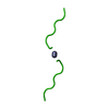
Journal: Nat Struct Mol Biol / Year: 2018
Title: Atomic structures of FUS LC domain segments reveal bases for reversible amyloid fibril formation.
Authors: Feng Luo / Xinrui Gui / Heng Zhou / Jinge Gu / Yichen Li / Xiangyu Liu / Minglei Zhao / Dan Li / Xueming Li / Cong Liu /
Abstract: Thermostable cross-β structures are characteristic of pathological amyloid fibrils, but these structures cannot explain the reversible nature of fibrils formed by RNA-binding proteins such as fused ...Thermostable cross-β structures are characteristic of pathological amyloid fibrils, but these structures cannot explain the reversible nature of fibrils formed by RNA-binding proteins such as fused in sarcoma (FUS), involved in RNA granule assembly. Here, we find that two tandem (S/G)Y(S/G) motifs of the human FUS low-complexity domain (FUS LC) form reversible fibrils in a temperature- and phosphorylation-dependent manner. We named these motifs reversible amyloid cores, or RAC1 and RAC2, and determined their atomic structures in fibrillar forms, using microelectron and X-ray diffraction techniques. The RAC1 structure features an ordered-coil fibril spine rather than the extended β-strand typical of amyloids. Ser42, a phosphorylation site of FUS, is critical in the maintenance of the ordered-coil structure, which explains how phosphorylation controls fibril formation. The RAC2 structure shows a labile fibril spine with a wet interface. These structures illuminate the mechanism of reversible fibril formation and dynamic assembly of RNA granules.
History
DepositionJun 9, 2017Deposition site: PDBJ / Processing site: PDBJ
Revision 1.0Apr 4, 2018Provider: repository / Type: Initial release
Revision 1.1Apr 18, 2018Group: Data collection / Database references / Category: citation
Item: _citation.journal_abbrev / _citation.journal_volume ..._citation.journal_abbrev / _citation.journal_volume / _citation.page_first / _citation.page_last / _citation.pdbx_database_id_PubMed / _citation.title
Revision 1.2Mar 27, 2024Group: Data collection / Database references / Derived calculations
Category: chem_comp_atom / chem_comp_bond ...chem_comp_atom / chem_comp_bond / database_2 / pdbx_struct_conn_angle / struct_conn
Item: _database_2.pdbx_DOI / _database_2.pdbx_database_accession ..._database_2.pdbx_DOI / _database_2.pdbx_database_accession / _pdbx_struct_conn_angle.ptnr1_auth_comp_id / _pdbx_struct_conn_angle.ptnr1_auth_seq_id / _pdbx_struct_conn_angle.ptnr1_label_atom_id / _pdbx_struct_conn_angle.ptnr1_label_comp_id / _pdbx_struct_conn_angle.ptnr1_label_seq_id / _pdbx_struct_conn_angle.ptnr2_symmetry / _pdbx_struct_conn_angle.ptnr3_auth_comp_id / _pdbx_struct_conn_angle.ptnr3_auth_seq_id / _pdbx_struct_conn_angle.ptnr3_label_atom_id / _pdbx_struct_conn_angle.ptnr3_label_comp_id / _pdbx_struct_conn_angle.ptnr3_label_seq_id / _pdbx_struct_conn_angle.value / _struct_conn.pdbx_dist_value / _struct_conn.ptnr1_auth_comp_id / _struct_conn.ptnr1_auth_seq_id / _struct_conn.ptnr1_label_atom_id / _struct_conn.ptnr1_label_comp_id / _struct_conn.ptnr1_label_seq_id / _struct_conn.ptnr2_symmetry

-
Structure visualization

Structure viewerMolecule:
MolmilJmol/JSmol

Downloads & links

-
Assembly

Deposited unit
A: RNA-binding protein FUS
hetero molecules


Theoretical massNumber of molelcules
Total (without water)7282
Polymers6631
Non-polymers651
Water905
1


  • Idetical with deposited unit
  • defined by author
  • Evidence: mass spectrometry
TypeNameSymmetry operationNumber
identity operation1_555x,y,z1
Buried area30 Å2
ΔGint-8 kcal/mol
Surface area940 Å2
2
A: RNA-binding protein FUS
hetero molecules

A: RNA-binding protein FUS
hetero molecules


Theoretical massNumber of molelcules
Total (without water)1,4564
Polymers1,3252
Non-polymers1312
Water362
TypeNameSymmetry operationNumber
identity operation1_555x,y,z1
crystal symmetry operation2_585-x,-y+3,z1
Buried area210 Å2
ΔGint-53 kcal/mol
Surface area1740 Å2
MethodPISA
Unit cell
Length a, b, c (Å)28.240, 30.270, 4.780
Angle α, β, γ (deg.)90.000, 90.000, 90.000
Int Tables number18
Space group name H-MP21212
Components on special symmetry positions
IDModelComponents
11A-201-

HOH

-
Components

#1: Protein/peptide RNA-binding protein FUS / 75 kDa DNA-pairing protein / Oncogene FUS / Oncogene TLS / POMp75 / Translocated in liposarcoma protein


Mass: 662.648 Da / Num. of mol.: 1 / Fragment: UNP RESIDUES 54-59 / Source method: obtained synthetically / Source: (synth.) Homo sapiens (human) / References: UniProt: P35637
#2: Chemical ChemComp-ZN / ZINC ION


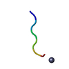
Mass: 65.409 Da / Num. of mol.: 1 / Source method: obtained synthetically / Formula: Zn
#3: Water ChemComp-HOH / water


Mass: 18.015 Da / Num. of mol.: 5 / Source method: isolated from a natural source / Formula: H2O

-
Experimental details

-
Experiment

ExperimentMethod: X-RAY DIFFRACTION / Number of used crystals: 1

-
Sample preparation

CrystalDensity Matthews: 1.54 Å3/Da / Density % sol: 20.21 %
Crystal growTemperature: 289 K / Method: vapor diffusion, hanging drop / pH: 8
Details: 2.5 M NaCl, 0.1 M imidazole pH 8.0, 0.2 M zinc acetate

-
Data collection

DiffractionMean temperature: 100 K
Diffraction sourceSource: SYNCHROTRON / Site: SSRF / Beamline: BL17U1 / Wavelength: 0.9791 Å
DetectorType: RAYONIX MX-225 / Detector: CCD / Date: Oct 28, 2015
RadiationProtocol: SINGLE WAVELENGTH / Monochromatic (M) / Laue (L): M / Scattering type: x-ray
Radiation wavelengthWavelength: 0.9791 Å / Relative weight: 1
ReflectionResolution: 1.5→20.649 Å / Num. obs: 1197 / % possible obs: 94.3 % / Observed criterion σ(I): -3 / Redundancy: 7.674 % / Biso Wilson estimate: 7.77 Å2 / CC1/2: 0.972 / Rmerge(I) obs: 0.422 / Rrim(I) all: 0.453 / Net I/σ(I): 3.37 / Num. measured all: 9186 / Scaling rejects: 0
Reflection shell

Diffraction-ID: 1

Resolution (Å)Redundancy (%)Rmerge(I) obsMean I/σ(I) obsNum. unique obsCC1/2Rrim(I) all% possible all
1.5-1.67.3450.8211.521940.7110.88175.8
1.6-1.87.7160.6272.422710.7980.673100
1.8-27.7730.5153.411980.8010.552100
2-2.37.8250.4833.982060.8240.517100
2.3-37.6770.4054.281640.9520.43493.7
3-57.7680.3145.411380.9790.339100
5-87.2940.3324.92170.990.362100
8-1080.3166.730.338100
10-20.6496.6670.2486.1760.9980.27100

-
Processing

Software
NameVersionClassification
XSCALEdata scaling
PHENIX1.11.1_2575refinement
PDB_EXTRACT3.22data extraction
RefinementResolution: 1.503→20.649 Å / SU ML: 0.17 / Cross valid method: FREE R-VALUE / σ(F): 1.44 / Phase error: 11.97
RfactorNum. reflection% reflection
Rfree0.2407 78 10.01 %
Rwork0.204 --
obs0.2098 779 94.88 %
Solvent computationShrinkage radii: 0.9 Å / VDW probe radii: 1.11 Å
Displacement parametersBiso max: 35.23 Å2 / Biso mean: 7.804 Å2 / Biso min: 1.16 Å2
Refinement stepCycle: final / Resolution: 1.503→20.649 Å
ProteinNucleic acidLigandSolventTotal
Num. atoms47 0 1 5 53
Biso mean--13.59 17.7 -
Num. residues----6
Refine LS restraints
Refine-IDTypeDev idealNumber
X-RAY DIFFRACTIONf_bond_d0.00748
X-RAY DIFFRACTIONf_angle_d1.1364
X-RAY DIFFRACTIONf_chiral_restr0.0615
X-RAY DIFFRACTIONf_plane_restr0.0058
X-RAY DIFFRACTIONf_dihedral_angle_d7.00525
LS refinement shellResolution: 1.5029→1.6 Å / Rfactor Rfree error: 0 / Total num. of bins used: 1
RfactorNum. reflection% reflection
Rfree0.2407 78 -
Rwork0.204 701 -
all-779 -
obs--95 %

+
About Yorodumi

-
News

-
Feb 9, 2022. New format data for meta-information of EMDB entries

New format data for meta-information of EMDB entries

  • Version 3 of the EMDB header file is now the official format.
  • The previous official version 1.9 will be removed from the archive.

Related info.:EMDB header

External links:wwPDB to switch to version 3 of the EMDB data model

-
Aug 12, 2020. Covid-19 info

Covid-19 info

URL: https://pdbjlvh1.pdbj.org/emnavi/covid19.php

New page: Covid-19 featured information page in EM Navigator.

Related info.:Covid-19 info / Mar 5, 2020. Novel coronavirus structure data

+
Mar 5, 2020. Novel coronavirus structure data

Novel coronavirus structure data

Related info.:Yorodumi Speices / Aug 12, 2020. Covid-19 info

External links:COVID-19 featured content - PDBj / Molecule of the Month (242):Coronavirus Proteases

+
Jan 31, 2019. EMDB accession codes are about to change! (news from PDBe EMDB page)

EMDB accession codes are about to change! (news from PDBe EMDB page)

  • The allocation of 4 digits for EMDB accession codes will soon come to an end. Whilst these codes will remain in use, new EMDB accession codes will include an additional digit and will expand incrementally as the available range of codes is exhausted. The current 4-digit format prefixed with “EMD-” (i.e. EMD-XXXX) will advance to a 5-digit format (i.e. EMD-XXXXX), and so on. It is currently estimated that the 4-digit codes will be depleted around Spring 2019, at which point the 5-digit format will come into force.
  • The EM Navigator/Yorodumi systems omit the EMD- prefix.

Related info.:Q: What is EMD? / ID/Accession-code notation in Yorodumi/EM Navigator

External links:EMDB Accession Codes are Changing Soon! / Contact to PDBj

+
Jul 12, 2017. Major update of PDB

Major update of PDB

  • wwPDB released updated PDB data conforming to the new PDBx/mmCIF dictionary.
  • This is a major update changing the version number from 4 to 5, and with Remediation, in which all the entries are updated.
  • In this update, many items about electron microscopy experimental information are reorganized (e.g. em_software).
  • Now, EM Navigator and Yorodumi are based on the updated data.

External links:wwPDB Remediation / Enriched Model Files Conforming to OneDep Data Standards Now Available in the PDB FTP Archive

-
Yorodumi

Thousand views of thousand structures

  • Yorodumi is a browser for structure data from EMDB, PDB, SASBDB, etc.
  • This page is also the successor to EM Navigator detail page, and also detail information page/front-end page for Omokage search.
  • The word "yorodu" (or yorozu) is an old Japanese word meaning "ten thousand". "mi" (miru) is to see.

Related info.:EMDB / PDB / SASBDB / Comparison of 3 databanks / Yorodumi Search / Aug 31, 2016. New EM Navigator & Yorodumi / Yorodumi Papers / Jmol/JSmol / Function and homology information / Changes in new EM Navigator and Yorodumi

Read more