 Movie
Movie Controller

[English] 日本語
 Yorodumi
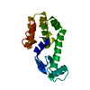
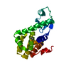
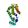
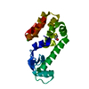
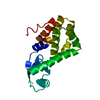
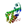
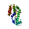
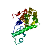
- PDB-5xpf: High-resolution X-ray structure of the T26H mutant of T4 lysozyme -
+ Open data

- Basic information
| Entry | Database: PDB / ID: 5xpf | |||||||||
|---|---|---|---|---|---|---|---|---|---|---|
| Title | High-resolution X-ray structure of the T26H mutant of T4 lysozyme | |||||||||
 Components Components | Endolysin | |||||||||
 Keywords Keywords | HYDROLASE / Alpha and beta protein / T26H mutant / Perdeuterated protein | |||||||||
| Function / homology |  Function and homology information Function and homology informationviral release from host cell by cytolysis / peptidoglycan catabolic process / cell wall macromolecule catabolic process / lysozyme / lysozyme activity / host cell cytoplasm / defense response to bacterium Similarity search - Function | |||||||||
| Biological species |  Enterobacteria phage T4 (virus) Enterobacteria phage T4 (virus) | |||||||||
| Method |  X-RAY DIFFRACTION / SYNCHROTRON / MOLECULAR REPLACEMENT / Resolution: 1.04 Å X-RAY DIFFRACTION / SYNCHROTRON / MOLECULAR REPLACEMENT / Resolution: 1.04 Å | |||||||||
 Authors Authors | Hiromoto, T. / Kuroki, R. | |||||||||
 Citation Citation | Journal: Protein Sci. / Year: 2017 Title: Neutron structure of the T26H mutant of T4 phage lysozyme provides insight into the catalytic activity of the mutant enzyme and how it differs from that of wild type. Authors: Hiromoto, T. / Meilleur, F. / Shimizu, R. / Shibazaki, C. / Adachi, M. / Tamada, T. / Kuroki, R. | |||||||||
| History |
|
- Structure visualization
| Structure viewer | Molecule: MolmilJmol/JSmol |
|---|
- Downloads & links
-Download
| PDBx/mmCIF format | 5xpf.cif.gz | 104.9 KB | Display | PDBx/mmCIF format |
|---|---|---|---|---|
| PDB format | pdb5xpf.ent.gz | 79.3 KB | Display | PDB format |
| PDBx/mmJSON format | 5xpf.json.gz | Tree view | PDBx/mmJSON format | |
| Others |  Other downloads Other downloads |
-Validation report
| Arichive directory | https://data.pdbj.org/pub/pdb/validation_reports/xp/5xpfftp://data.pdbj.org/pub/pdb/validation_reports/xp/5xpf | HTTPS FTP |
|---|
-Related structure data
| Related structure data |  5xpeC  1qt8S S: Starting model for refinement C: citing same article ( |
|---|---|
| Similar structure data |


-Links
 PDBj
PDBj


- Assembly
| Deposited unit | 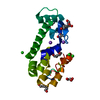
| ||||||||
|---|---|---|---|---|---|---|---|---|---|
| 1 |
| ||||||||
| Unit cell |
|
-Components
-Protein , 1 types, 1 molecules A
| #1: Protein | Mass: 18665.406 Da / Num. of mol.: 1 / Mutation: T26H, C54T, C97A Source method: isolated from a genetically manipulated source Source: (gene. exp.) Enterobacteria phage T4 (virus) / Gene: e, T4Tp126 / Plasmid: pET24 / Production host:  |
|---|
-Non-polymers , 5 types, 257 molecules 


| #2: Chemical |  Mass: 22.990 Da / Num. of mol.: 3 / Source method: obtained synthetically / Formula: Na Mass: 22.990 Da / Num. of mol.: 3 / Source method: obtained synthetically / Formula: Na#3: Chemical | ChemComp-CL /  Mass: 35.453 Da / Num. of mol.: 5 / Source method: obtained synthetically / Formula: Cl Mass: 35.453 Da / Num. of mol.: 5 / Source method: obtained synthetically / Formula: Cl#4: Chemical | ChemComp-HEZ /  Mass: 118.174 Da / Num. of mol.: 4 / Source method: obtained synthetically / Formula: C6H14O2 Mass: 118.174 Da / Num. of mol.: 4 / Source method: obtained synthetically / Formula: C6H14O2#5: Chemical |  Mass: 92.094 Da / Num. of mol.: 3 / Source method: obtained synthetically / Formula: C3H8O3 Mass: 92.094 Da / Num. of mol.: 3 / Source method: obtained synthetically / Formula: C3H8O3#6: Water | ChemComp-HOH / | Mass: 18.015 Da / Num. of mol.: 242 / Source method: isolated from a natural source / Formula: H2O |
|---|
-Experimental details
-Experiment
| Experiment | Method: X-RAY DIFFRACTION / Number of used crystals: 1 |
|---|
- Sample preparation
| Crystal | Density Matthews: 2.65 Å3/Da / Density % sol: 53.64 % |
|---|---|
| Crystal grow | Temperature: 293 K / Method: vapor diffusion, sitting drop / pH: 7 Details: Sodium-potassium phosphate, Sodium chloride, 1,6-Hexanediol |
-Data collection
| Diffraction | Mean temperature: 100 K |
|---|---|
| Diffraction source | Source: SYNCHROTRON / Site: Photon Factory  / Beamline: BL-5A / Wavelength: 0.8 Å / Beamline: BL-5A / Wavelength: 0.8 Å |
| Detector | Type: ADSC QUANTUM 315r / Detector: CCD / Date: Nov 30, 2014 |
| Radiation | Monochromator: Si(111) double crystal / Protocol: SINGLE WAVELENGTH / Monochromatic (M) / Laue (L): M / Scattering type: x-ray |
| Radiation wavelength | Wavelength: 0.8 Å / Relative weight: 1 |
| Reflection | Resolution: 1.04→50 Å / Num. obs: 95774 / % possible obs: 99.9 % / Redundancy: 4.4 % / Rmerge(I) obs: 0.041 / Rpim(I) all: 0.019 / Rrim(I) all: 0.045 / Rsym value: 0.041 / Χ2: 2.263 / Net I/av σ(I): 51.6 / Net I/σ(I): 51.6 |
| Reflection shell | Resolution: 1.04→1.08 Å / Redundancy: 3.7 % / Rmerge(I) obs: 0.361 / Mean I/σ(I) obs: 4.7 / Num. unique obs: 9481 / CC1/2: 0.886 / Rpim(I) all: 0.215 / Rrim(I) all: 0.422 / Rsym value: 0.361 / Χ2: 1.732 / % possible all: 100 |
- Processing
| Software |
| |||||||||||||||||||||||||||||||||
|---|---|---|---|---|---|---|---|---|---|---|---|---|---|---|---|---|---|---|---|---|---|---|---|---|---|---|---|---|---|---|---|---|---|---|
| Refinement | Method to determine structure: MOLECULAR REPLACEMENT Starting model: 1QT8 Resolution: 1.04→45.58 Å / Cross valid method: FREE R-VALUE / σ(F): 0 / Details: ANISOTROPIC REFINEMENT REDUCED FREE R
| |||||||||||||||||||||||||||||||||
| Displacement parameters | Biso max: 96.95 Å2 / Biso mean: 18.1199 Å2 / Biso min: 7.69 Å2 | |||||||||||||||||||||||||||||||||
| Refinement step | Cycle: final / Resolution: 1.04→45.58 Å
| |||||||||||||||||||||||||||||||||
| Refine LS restraints |
|
