 Movie
Movie Controller

[English] 日本語
 Yorodumi
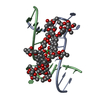
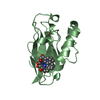
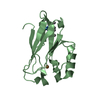
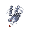
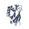
- PDB-5xjw: Crystal Structure of the [Co2+-(Chromomycin A3)2]-CCG repeats Complex -
+ Open data

- Basic information
| Entry | Database: PDB / ID: 5xjw | |||||||||||||||||||||||||||||
|---|---|---|---|---|---|---|---|---|---|---|---|---|---|---|---|---|---|---|---|---|---|---|---|---|---|---|---|---|---|---|
| Title | Crystal Structure of the [Co2+-(Chromomycin A3)2]-CCG repeats Complex | |||||||||||||||||||||||||||||
 Components Components | DNA (5'-D(* Keywords KeywordsDNA/ANTIBIOTIC / Nucleotide Flipping-out / CCG trinucleotide repeats / Chromomycin A3 / Metalloantibiotics / Minor Groove Binding drug / DNA-ANTIBIOTIC complex | Function / homology | Chromomycin A3, Monomer / : / Chem-CPH / DNA / DNA (> 10) |  Function and homology information Function and homology informationBiological species | synthetic construct (others) | Method |  X-RAY DIFFRACTION / SYNCHROTRON / MOLECULAR REPLACEMENT / Resolution: 2.097 Å X-RAY DIFFRACTION / SYNCHROTRON / MOLECULAR REPLACEMENT / Resolution: 2.097 Å  Authors AuthorsTseng, W.H. / Wu, P.C. / Satange, R.B. / Hou, M.H. |  CitationJournal: To be published CitationJournal: To be publishedTitle: Crystal Structure of the [Co2+-(Chromomycin A3)2]-CCG repeats Complex Authors: Tseng, W.H. / Wu, P.C. / Satange, R.B. / Hou, M.H. History |
|
- Structure visualization
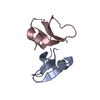
| Structure viewer | Molecule: MolmilJmol/JSmol |
|---|
- Downloads & links
-Download
| PDBx/mmCIF format | 5xjw.cif.gz | 34.5 KB | Display | PDBx/mmCIF format |
|---|---|---|---|---|
| PDB format | pdb5xjw.ent.gz | 21 KB | Display | PDB format |
| PDBx/mmJSON format | 5xjw.json.gz | Tree view | PDBx/mmJSON format | |
| Others |  Other downloads Other downloads |
-Validation report
| Arichive directory | https://data.pdbj.org/pub/pdb/validation_reports/xj/5xjwftp://data.pdbj.org/pub/pdb/validation_reports/xj/5xjw | HTTPS FTP |
|---|
-Related structure data
| Related structure data |  5xewS S: Starting model for refinement |
|---|---|
| Similar structure data |

-Links
 PDBj
PDBj


- Assembly
| Deposited unit | 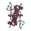
| ||||||||
|---|---|---|---|---|---|---|---|---|---|
| 1 |
| ||||||||
| Unit cell |
| ||||||||
| Components on special symmetry positions |
|
-Components
-DNA chain , 1 types, 2 molecules AB
| #1: DNA chain | Mass: 3912.550 Da / Num. of mol.: 2 / Source method: obtained synthetically / Source: (synth.) synthetic construct (others) |
|---|
-Sugars , 2 types, 4 molecules
| #2: Polysaccharide | 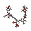  Source method: isolated from a genetically manipulated source References: Chromomycin A3, Monomer #3: Polysaccharide | Source method: isolated from a genetically manipulated source References: Chromomycin A3, Monomer |
|---|
-Non-polymers , 3 types, 55 molecules 


| #4: Chemical |  Type: Oligosaccharide / Class: Antibiotic / Mass: 420.410 Da / Num. of mol.: 2 / Source method: obtained synthetically / Formula: C21H24O9 / References: Chromomycin A3, Monomer Type: Oligosaccharide / Class: Antibiotic / Mass: 420.410 Da / Num. of mol.: 2 / Source method: obtained synthetically / Formula: C21H24O9 / References: Chromomycin A3, Monomer#5: Chemical |  Mass: 58.933 Da / Num. of mol.: 3 / Source method: obtained synthetically / Formula: Co Mass: 58.933 Da / Num. of mol.: 3 / Source method: obtained synthetically / Formula: Co#6: Water | ChemComp-HOH / | Mass: 18.015 Da / Num. of mol.: 50 / Source method: isolated from a natural source / Formula: H2O |
|---|
-Experimental details
-Experiment
| Experiment | Method: X-RAY DIFFRACTION / Number of used crystals: 1 |
|---|
- Sample preparation
| Crystal | Density Matthews: 3.89 Å3/Da / Density % sol: 68.36 % |
|---|---|
| Crystal grow | Temperature: 277 K / Method: vapor diffusion, sitting drop / pH: 6 Details: 1.0 mM ssDNA, 2.0 mM Chromomycin A3, 4mM CoCl2.6H2O, 50 mM Sodium Cacodylate, 5 mM Spermine, 3% MPD |
-Data collection
| Diffraction | Mean temperature: 100 K | ||||||||||||||||||||||||||||||||||||||||||||||||||||||||||||||||||
|---|---|---|---|---|---|---|---|---|---|---|---|---|---|---|---|---|---|---|---|---|---|---|---|---|---|---|---|---|---|---|---|---|---|---|---|---|---|---|---|---|---|---|---|---|---|---|---|---|---|---|---|---|---|---|---|---|---|---|---|---|---|---|---|---|---|---|---|
| Diffraction source | Source: SYNCHROTRON / Site: NSRRC  / Beamline: BL13B1 / Wavelength: 1.56418 Å / Beamline: BL13B1 / Wavelength: 1.56418 Å | ||||||||||||||||||||||||||||||||||||||||||||||||||||||||||||||||||
| Detector | Type: ADSC QUANTUM 315r / Detector: CCD / Date: Sep 18, 2012 | ||||||||||||||||||||||||||||||||||||||||||||||||||||||||||||||||||
| Radiation | Protocol: SINGLE WAVELENGTH / Monochromatic (M) / Laue (L): M / Scattering type: x-ray | ||||||||||||||||||||||||||||||||||||||||||||||||||||||||||||||||||
| Radiation wavelength | Wavelength: 1.56418 Å / Relative weight: 1 | ||||||||||||||||||||||||||||||||||||||||||||||||||||||||||||||||||
| Reflection | Resolution: 2.097→30 Å / Num. obs: 12596 / % possible obs: 99.6 % / Redundancy: 6.1 % / Biso Wilson estimate: 47.68 Å2 / Rmerge(I) obs: 0.055 / Χ2: 1.887 / Net I/σ(I): 24.8 | ||||||||||||||||||||||||||||||||||||||||||||||||||||||||||||||||||
| Reflection shell |
|
- Processing
| Software |
| ||||||||||||||||||||||||||||||||||||||||||||||||||||||||||||||||||||||
|---|---|---|---|---|---|---|---|---|---|---|---|---|---|---|---|---|---|---|---|---|---|---|---|---|---|---|---|---|---|---|---|---|---|---|---|---|---|---|---|---|---|---|---|---|---|---|---|---|---|---|---|---|---|---|---|---|---|---|---|---|---|---|---|---|---|---|---|---|---|---|---|
| Refinement | Method to determine structure: MOLECULAR REPLACEMENT Starting model: 5XEW Resolution: 2.097→29.576 Å / SU ML: 0.25 / Cross valid method: THROUGHOUT / σ(F): 1.36 / Phase error: 31.07 Details: The force field of Chro unit used in the refinement of CoII(Chro)2-DNA complex structure was generated using the atomic resolution crystal structure of NiII(Chro)2.
| ||||||||||||||||||||||||||||||||||||||||||||||||||||||||||||||||||||||
| Solvent computation | Shrinkage radii: 0.9 Å / VDW probe radii: 1.11 Å | ||||||||||||||||||||||||||||||||||||||||||||||||||||||||||||||||||||||
| Displacement parameters | Biso max: 123.08 Å2 / Biso mean: 55.6101 Å2 / Biso min: 30.93 Å2 | ||||||||||||||||||||||||||||||||||||||||||||||||||||||||||||||||||||||
| Refinement step | Cycle: final / Resolution: 2.097→29.576 Å
| ||||||||||||||||||||||||||||||||||||||||||||||||||||||||||||||||||||||
| Refine LS restraints |
| ||||||||||||||||||||||||||||||||||||||||||||||||||||||||||||||||||||||
| LS refinement shell | Refine-ID: X-RAY DIFFRACTION / Rfactor Rfree error: 0 / Total num. of bins used: 9
|
