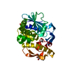
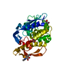
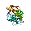
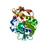
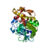
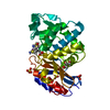
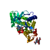
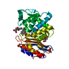
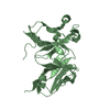
[English] 日本語
Yorodumi
- PDB-5wv1: Crystal structure of the complex of Ribosome inactivating protein... -

+
Open data


ID or keywords:

Loading...

-
Basic information

Entry
Database: PDB / ID: 5wv1
TitleCrystal structure of the complex of Ribosome inactivating protein from Momordica balsamina with ribose sugar at 1.90 A resolution.
ComponentsRibosome inactivating proteinRibosome-inactivating protein
KeywordsHYDROLASE
Function / homology
Function and homology information


rRNA N-glycosylase / rRNA N-glycosylase activity / toxin activity / negative regulation of translation / nucleotide binding
Similarity search - Function
Ricin (A Subunit), domain 2 / Ricin (A Subunit), domain 2 / Ricin (A subunit); domain 1 / Ricin (A subunit), domain 1 / Ribosome-inactivating protein conserved site / Shiga/ricin ribosomal inactivating toxins active site signature. / Ribosome-inactivating protein type 1/2 / Ribosome-inactivating protein / Ribosome-inactivating protein, subdomain 1 / Ribosome-inactivating protein, subdomain 2 ...Ricin (A Subunit), domain 2 / Ricin (A Subunit), domain 2 / Ricin (A subunit); domain 1 / Ricin (A subunit), domain 1 / Ribosome-inactivating protein conserved site / Shiga/ricin ribosomal inactivating toxins active site signature. / Ribosome-inactivating protein type 1/2 / Ribosome-inactivating protein / Ribosome-inactivating protein, subdomain 1 / Ribosome-inactivating protein, subdomain 2 / Ribosome-inactivating protein superfamily / Ribosome inactivating protein / Few Secondary Structures / Irregular / 3-Layer(aba) Sandwich / Alpha Beta
Similarity search - Domain/homology
alpha-D-ribofuranose / rRNA N-glycosylase
Similarity search - Component
Biological speciesMomordica balsamina (balsam apple)
MethodX-RAY DIFFRACTION / SYNCHROTRON / MOLECULAR REPLACEMENT / Resolution: 1.9 Å
AuthorsShokeen, A. / Singh, P.K. / Pandey, S. / Kaur, P. / Sharma, S. / Singh, T.P.
CitationJournal: To Be Published
Title: Crystal structure of the complex of Ribosome inactivating protein from Momordica balsamina with ribose sugar at 1.90 A resolution.
Authors: Shokeen, A. / Singh, P.K. / Pandey, S.
History
DepositionDec 21, 2016Deposition site: PDBJ / Processing site: PDBJ
Revision 1.0Jan 25, 2017Provider: repository / Type: Initial release
Revision 2.0Jul 29, 2020Group: Atomic model / Data collection ...Atomic model / Data collection / Derived calculations / Refinement description / Structure summary
Category: atom_site / chem_comp ...atom_site / chem_comp / entity / pdbx_chem_comp_identifier / pdbx_entity_nonpoly / refine_hist / struct_conn / struct_site / struct_site_gen
Item: _atom_site.auth_atom_id / _atom_site.label_atom_id ..._atom_site.auth_atom_id / _atom_site.label_atom_id / _chem_comp.name / _chem_comp.type / _entity.pdbx_description / _pdbx_entity_nonpoly.name / _refine_hist.d_res_low / _struct_conn.pdbx_role
Description: Carbohydrate remediation / Provider: repository / Type: Remediation
Revision 2.1Nov 22, 2023Group: Data collection / Database references ...Data collection / Database references / Refinement description / Structure summary
Category: chem_comp / chem_comp_atom ...chem_comp / chem_comp_atom / chem_comp_bond / database_2 / pdbx_initial_refinement_model
Item: _chem_comp.pdbx_synonyms / _database_2.pdbx_DOI / _database_2.pdbx_database_accession

-
Structure visualization

Structure viewerMolecule:
MolmilJmol/JSmol

Downloads & links

-
Assembly

Deposited unit
A: Ribosome inactivating protein
hetero molecules


Theoretical massNumber of molelcules
Total (without water)27,5574
Polymers27,0941
Non-polymers4633
Water3,333185
1


  • Idetical with deposited unit
  • defined by author
TypeNameSymmetry operationNumber
identity operation1_555x,y,z1
Unit cell
Length a, b, c (Å)129.748, 129.748, 41.457
Angle α, β, γ (deg.)90.00, 90.00, 120.00
Int Tables number146
Space group name H-MH3

-
Components

#1: Protein Ribosome inactivating protein / Ribosome-inactivating protein


Mass: 27093.756 Da / Num. of mol.: 1 / Source method: isolated from a natural source / Source: (natural) Momordica balsamina (balsam apple) / References: UniProt: D9J2T9, rRNA N-glycosylase
#2: Sugar ChemComp-NAG / 2-acetamido-2-deoxy-beta-D-glucopyranose / N-acetyl-beta-D-glucosamine / 2-acetamido-2-deoxy-beta-D-glucose / 2-acetamido-2-deoxy-D-glucose / 2-acetamido-2-deoxy-glucose / N-ACETYL-D-GLUCOSAMINE / N-Acetylglucosamine


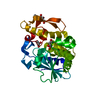
Type: D-saccharide, beta linking / Mass: 221.208 Da / Num. of mol.: 1
Source method: isolated from a genetically manipulated source
Formula: C8H15NO6
IdentifierTypeProgram
DGlcpNAcbCONDENSED IUPAC CARBOHYDRATE SYMBOLGMML 1.0
N-acetyl-b-D-glucopyranosamineCOMMON NAMEGMML 1.0
b-D-GlcpNAcIUPAC CARBOHYDRATE SYMBOLPDB-CARE 1.0
GlcNAcSNFG CARBOHYDRATE SYMBOLGMML 1.0
#3: Chemical ChemComp-GOL / GLYCEROL / GLYCERIN / PROPANE-1,2,3-TRIOL / Glycerol


Mass: 92.094 Da / Num. of mol.: 1 / Source method: obtained synthetically / Formula: C3H8O3
#4: Sugar ChemComp-RIB / alpha-D-ribofuranose / alpha-D-ribose / D-ribose / ribose / Ribose


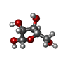
Type: D-saccharide, alpha linking / Mass: 150.130 Da / Num. of mol.: 1
Source method: isolated from a genetically manipulated source
Formula: C5H10O5
IdentifierTypeProgram
DRibfaCONDENSED IUPAC CARBOHYDRATE SYMBOLGMML 1.0
a-D-ribofuranoseCOMMON NAMEGMML 1.0
a-D-RibfIUPAC CARBOHYDRATE SYMBOLPDB-CARE 1.0
RibSNFG CARBOHYDRATE SYMBOLGMML 1.0
#5: Water ChemComp-HOH / water / Water


Mass: 18.015 Da / Num. of mol.: 185 / Source method: isolated from a natural source / Formula: H2O

-
Experimental details

-
Experiment

ExperimentMethod: X-RAY DIFFRACTION / Number of used crystals: 1

-
Sample preparation

CrystalDensity Matthews: 2.48 Å3/Da / Density % sol: 50.47 %
Crystal growTemperature: 298 K / Method: vapor diffusion, hanging drop / pH: 6.7
Details: 14% PEG 6000, 0.1M Sodium Phosphate, pH 6.7, VAPOR DIFFUSION, HANGING DROP, temperature 298K

-
Data collection

DiffractionMean temperature: 100 K
Diffraction sourceSource: SYNCHROTRON / Site: ESRF / Beamline: BM14 / Wavelength: 0.97 Å
DetectorType: MARRESEARCH / Detector: CCD / Date: Nov 28, 2015 / Details: MIRROR
RadiationMonochromator: GRAPHITE / Protocol: SINGLE WAVELENGTH / Monochromatic (M) / Laue (L): M / Scattering type: x-ray
Radiation wavelengthWavelength: 0.97 Å / Relative weight: 1
ReflectionResolution: 1.9→38.89 Å / Num. obs: 20400 / % possible obs: 99.4 % / Redundancy: 5.6 % / Rsym value: 0.09 / Net I/σ(I): 19.8
Reflection shellResolution: 1.9→1.93 Å / Redundancy: 4.5 % / Mean I/σ(I) obs: 1.23 / % possible all: 95.2

-
Processing

Software
NameVersionClassification
REFMAC5.8.0135refinement
HKL-2000data reduction
SCALEPACKdata scaling
MOLREPphasing
RefinementMethod to determine structure: MOLECULAR REPLACEMENT
Starting model: 3RL9
Resolution: 1.9→38.89 Å / Cor.coef. Fo:Fc: 0.975 / Cor.coef. Fo:Fc free: 0.951 / SU B: 8.518 / SU ML: 0.121 / Cross valid method: THROUGHOUT / ESU R: 0.131 / ESU R Free: 0.134 / Stereochemistry target values: MAXIMUM LIKELIHOOD / Details: HYDROGENS HAVE BEEN ADDED IN THE RIDING POSITIONS
RfactorNum. reflection% reflectionSelection details
Rfree0.20887 1007 4.9 %RANDOM
Rwork0.1531 ---
obs0.15588 19391 99.38 %-
Solvent computationIon probe radii: 0.8 Å / Shrinkage radii: 0.8 Å / VDW probe radii: 1.2 Å / Solvent model: MASK
Displacement parametersBiso mean: 34.155 Å2
Baniso -1Baniso -2Baniso -3
1--0.27 Å2-0.13 Å20 Å2
2---0.27 Å20 Å2
3---0.86 Å2
Refinement stepCycle: 1 / Resolution: 1.9→38.89 Å
ProteinNucleic acidLigandSolventTotal
Num. atoms1911 0 30 185 2126
Refine LS restraints
Refine-IDTypeDev idealDev ideal targetNumber
X-RAY DIFFRACTIONr_bond_refined_d0.0280.0191976
X-RAY DIFFRACTIONr_bond_other_d0.0040.021906
X-RAY DIFFRACTIONr_angle_refined_deg2.4461.9822690
X-RAY DIFFRACTIONr_angle_other_deg1.23734368
X-RAY DIFFRACTIONr_dihedral_angle_1_deg6.1725245
X-RAY DIFFRACTIONr_dihedral_angle_2_deg36.07923.92984
X-RAY DIFFRACTIONr_dihedral_angle_3_deg15.32815322
X-RAY DIFFRACTIONr_dihedral_angle_4_deg18.1731513
X-RAY DIFFRACTIONr_chiral_restr0.1460.2322
X-RAY DIFFRACTIONr_gen_planes_refined0.0120.0212224
X-RAY DIFFRACTIONr_gen_planes_other0.0020.02449
X-RAY DIFFRACTIONr_nbd_refined
X-RAY DIFFRACTIONr_nbd_other
X-RAY DIFFRACTIONr_nbtor_refined
X-RAY DIFFRACTIONr_nbtor_other
X-RAY DIFFRACTIONr_xyhbond_nbd_refined
X-RAY DIFFRACTIONr_xyhbond_nbd_other
X-RAY DIFFRACTIONr_metal_ion_refined
X-RAY DIFFRACTIONr_metal_ion_other
X-RAY DIFFRACTIONr_symmetry_vdw_refined
X-RAY DIFFRACTIONr_symmetry_vdw_other
X-RAY DIFFRACTIONr_symmetry_hbond_refined
X-RAY DIFFRACTIONr_symmetry_hbond_other
X-RAY DIFFRACTIONr_symmetry_metal_ion_refined
X-RAY DIFFRACTIONr_symmetry_metal_ion_other
X-RAY DIFFRACTIONr_mcbond_it1.1111.618983
X-RAY DIFFRACTIONr_mcbond_other1.1091.617982
X-RAY DIFFRACTIONr_mcangle_it1.542.4171227
X-RAY DIFFRACTIONr_mcangle_other1.542.4181228
X-RAY DIFFRACTIONr_scbond_it1.8771.902993
X-RAY DIFFRACTIONr_scbond_other1.8671.903993
X-RAY DIFFRACTIONr_scangle_it
X-RAY DIFFRACTIONr_scangle_other2.8242.7631464
X-RAY DIFFRACTIONr_long_range_B_refined6.32414.9172364
X-RAY DIFFRACTIONr_long_range_B_other6.32214.9342365
X-RAY DIFFRACTIONr_rigid_bond_restr
X-RAY DIFFRACTIONr_sphericity_free
X-RAY DIFFRACTIONr_sphericity_bonded
LS refinement shellResolution: 1.9→1.949 Å / Total num. of bins used: 20
RfactorNum. reflection% reflection
Rfree0.302 71 -
Rwork0.272 1384 -
obs--94.66 %
Refinement TLS params.Method: refined / Origin x: -20.3029 Å / Origin y: -12.6873 Å / Origin z: -13.3128 Å
111213212223313233
T0.0135 Å2-0.0102 Å2-0.0272 Å2-0.0144 Å20.0047 Å2--0.2762 Å2
L2.838 °20.2848 °2-0.8223 °2-1.7788 °2-0.3158 °2--0.9843 °2
S-0.04 Å °-0.0299 Å °-0.2476 Å °-0.0052 Å °0.0292 Å °0.2041 Å °0.0844 Å °-0.0741 Å °0.0108 Å °
Refinement TLS group
IDRefine-IDRefine TLS-IDAuth asym-IDAuth seq-ID
1X-RAY DIFFRACTION1A1 - 246
2X-RAY DIFFRACTION1A301

+
About Yorodumi

-
News

-
Feb 9, 2022. New format data for meta-information of EMDB entries

New format data for meta-information of EMDB entries

  • Version 3 of the EMDB header file is now the official format.
  • The previous official version 1.9 will be removed from the archive.

Related info.:EMDB header

External links:wwPDB to switch to version 3 of the EMDB data model

-
Aug 12, 2020. Covid-19 info

Covid-19 info

URL: https://pdbj.org/emnavi/covid19.php

New page: Covid-19 featured information page in EM Navigator.

Related info.:Covid-19 info / Mar 5, 2020. Novel coronavirus structure data

+
Mar 5, 2020. Novel coronavirus structure data

Novel coronavirus structure data

Related info.:Yorodumi Speices / Aug 12, 2020. Covid-19 info

External links:COVID-19 featured content - PDBj / Molecule of the Month (242):Coronavirus Proteases

+
Jan 31, 2019. EMDB accession codes are about to change! (news from PDBe EMDB page)

EMDB accession codes are about to change! (news from PDBe EMDB page)

  • The allocation of 4 digits for EMDB accession codes will soon come to an end. Whilst these codes will remain in use, new EMDB accession codes will include an additional digit and will expand incrementally as the available range of codes is exhausted. The current 4-digit format prefixed with “EMD-” (i.e. EMD-XXXX) will advance to a 5-digit format (i.e. EMD-XXXXX), and so on. It is currently estimated that the 4-digit codes will be depleted around Spring 2019, at which point the 5-digit format will come into force.
  • The EM Navigator/Yorodumi systems omit the EMD- prefix.

Related info.:Q: What is EMD? / ID/Accession-code notation in Yorodumi/EM Navigator

External links:EMDB Accession Codes are Changing Soon! / Contact to PDBj

+
Jul 12, 2017. Major update of PDB

Major update of PDB

  • wwPDB released updated PDB data conforming to the new PDBx/mmCIF dictionary.
  • This is a major update changing the version number from 4 to 5, and with Remediation, in which all the entries are updated.
  • In this update, many items about electron microscopy experimental information are reorganized (e.g. em_software).
  • Now, EM Navigator and Yorodumi are based on the updated data.

External links:wwPDB Remediation / Enriched Model Files Conforming to OneDep Data Standards Now Available in the PDB FTP Archive

-
Yorodumi

Thousand views of thousand structures

  • Yorodumi is a browser for structure data from EMDB, PDB, SASBDB, etc.
  • This page is also the successor to EM Navigator detail page, and also detail information page/front-end page for Omokage search.
  • The word "yorodu" (or yorozu) is an old Japanese word meaning "ten thousand". "mi" (miru) is to see.

Related info.:EMDB / PDB / SASBDB / Comparison of 3 databanks / Yorodumi Search / Aug 31, 2016. New EM Navigator & Yorodumi / Yorodumi Papers / Jmol/JSmol / Function and homology information / Changes in new EM Navigator and Yorodumi

Read more