| Entry | Database: PDB / ID: 5wph
|
|---|
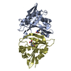
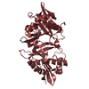
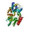
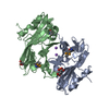
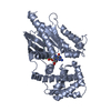
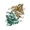
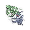
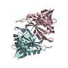
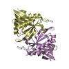
| Title | Crystal structure of ArsN, N-acetyltransferase with substrate AST from Pseudomonas putida KT2440 |
|---|
 Components Components | Phosphinothricin N-acetyltransferase |
|---|
 Keywords Keywords | TRANSFERASE / N-acetyltransferase / Pseudomonas putida |
|---|
| Function / homology |  Function and homology information Function and homology information
Acetyltransferase (GNAT) domain / Gcn5-related N-acetyltransferase (GNAT) / Gcn5-related N-acetyltransferase (GNAT) domain profile. / GNAT domain / Acyl-CoA N-acyltransferase / Aminopeptidase / 3-Layer(aba) Sandwich / Alpha BetaSimilarity search - Domain/homology |
|---|
| Biological species |  Pseudomonas putida (bacteria) Pseudomonas putida (bacteria) |
|---|
| Method |  X-RAY DIFFRACTION / SYNCHROTRON / MOLECULAR REPLACEMENT / Resolution: 2.19 Å X-RAY DIFFRACTION / SYNCHROTRON / MOLECULAR REPLACEMENT / Resolution: 2.19 Å |
|---|
 Authors Authors | Venkadesh, S. / Dheeman, D.S. / Yoshinaga, M. / Kandavelu, P. / Rosen, B.P. |
|---|
| Funding support |  United States, 1items United States, 1items | Organization | Grant number | Country |
|---|
| National Institutes of Health/National Institute of Environmental Health Sciences (NIH/NIEHS) | R37 GM55425 | United States |
|
|---|
 Citation Citation | Journal: Commun Biol / Year: 2019
Title: Arsinothricin, an arsenic-containing non-proteinogenic amino acid analog of glutamate, is a broad-spectrum antibiotic.
Authors: Nadar, V.S. / Chen, J. / Dheeman, D.S. / Galvan, A.E. / Yoshinaga-Sakurai, K. / Kandavelu, P. / Sankaran, B. / Kuramata, M. / Ishikawa, S. / Rosen, B.P. / Yoshinaga, M. |
|---|
| History | | Deposition | Aug 4, 2017 | Deposition site: RCSB / Processing site: RCSB |
|---|
| Revision 1.0 | Oct 10, 2018 | Provider: repository / Type: Initial release |
|---|
| Revision 1.1 | Apr 24, 2019 | Group: Data collection / Database references / Structure summary
Category: citation / citation_author / entity
Item: _citation.country / _citation.journal_abbrev ..._citation.country / _citation.journal_abbrev / _citation.journal_id_CSD / _citation.journal_id_ISSN / _citation.journal_volume / _citation.pdbx_database_id_DOI / _citation.title / _citation.year / _entity.formula_weight |
|---|
| Revision 1.2 | May 1, 2019 | Group: Data collection / Database references / Category: citation / citation_author
Item: _citation.page_first / _citation.page_last ..._citation.page_first / _citation.page_last / _citation.pdbx_database_id_PubMed / _citation.title / _citation_author.name |
|---|
| Revision 1.3 | Dec 18, 2019 | Group: Author supporting evidence / Category: pdbx_audit_support / Item: _pdbx_audit_support.funding_organization |
|---|
| Revision 1.4 | Oct 4, 2023 | Group: Data collection / Database references / Refinement description
Category: chem_comp_atom / chem_comp_bond ...chem_comp_atom / chem_comp_bond / database_2 / pdbx_initial_refinement_model
Item: _database_2.pdbx_DOI / _database_2.pdbx_database_accession |
|---|
|
|---|
 Movie
Movie Controller

 Yorodumi
 Open data
 Basic information
 Structure visualization
 Downloads & links
 Other downloads

 PDBj
PDBj

 Assembly
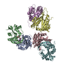
 Mass: 225.075 Da / Num. of mol.: 2 / Source method: obtained synthetically / Formula: C5H12AsNO4
 Mass: 22.990 Da / Num. of mol.: 4 / Source method: obtained synthetically / Formula: Na
 Mass: 18.015 Da / Num. of mol.: 875 / Source method: isolated from a natural source / Formula: H2O
 Sample preparation
 Processing