 Movie
Movie Controller

[English] 日本語
 Yorodumi
- PDB-5w8d: The structure of a COA-dependent acyl-homoserine lactone synthase... -
+ Open data

- Basic information
| Entry | Database: PDB / ID: 5w8d | ||||||
|---|---|---|---|---|---|---|---|
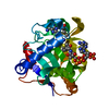
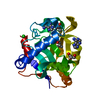
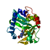
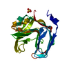
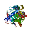
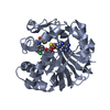
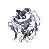
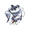
| Title | The structure of a COA-dependent acyl-homoserine lactone synthase, BjaI, with MTA | ||||||
 Components Components | Autoinducer synthase | ||||||
 Keywords Keywords | BIOSYNTHETIC PROTEIN / acyl-homoserine lactone / coenzyme A / BjaI | ||||||
| Function / homology | 5'-DEOXY-5'-METHYLTHIOADENOSINE / :  Function and homology information Function and homology information | ||||||
| Biological species |  Bradyrhizobium japonicum (bacteria) Bradyrhizobium japonicum (bacteria) | ||||||
| Method |  X-RAY DIFFRACTION / SYNCHROTRON / MAD / Resolution: 2 Å X-RAY DIFFRACTION / SYNCHROTRON / MAD / Resolution: 2 Å | ||||||
 Authors Authors | Dong, S.-H. / Nair, S.K. | ||||||
 Citation Citation | Journal: Proc. Natl. Acad. Sci. U.S.A. / Year: 2017 Title: Molecular basis for the substrate specificity of quorum signal synthases. Authors: Dong, S.H. / Frane, N.D. / Christensen, Q.H. / Greenberg, E.P. / Nagarajan, R. / Nair, S.K. | ||||||
| History |
|
- Structure visualization
| Structure viewer | Molecule: MolmilJmol/JSmol |
|---|
- Downloads & links
-Download
| PDBx/mmCIF format | 5w8d.cif.gz | 64.1 KB | Display | PDBx/mmCIF format |
|---|---|---|---|---|
| PDB format | pdb5w8d.ent.gz | 45.4 KB | Display | PDB format |
| PDBx/mmJSON format | 5w8d.json.gz | Tree view | PDBx/mmJSON format | |
| Others |  Other downloads Other downloads |
-Validation report
| Arichive directory | https://data.pdbj.org/pub/pdb/validation_reports/w8/5w8dftp://data.pdbj.org/pub/pdb/validation_reports/w8/5w8d | HTTPS FTP |
|---|
-Related structure data


-Links
 PDBj
PDBj- Assembly
| Deposited unit | 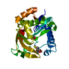
| ||||||||
|---|---|---|---|---|---|---|---|---|---|
| 1 |
| ||||||||
| Unit cell |
|
-Components
| #1: Protein | Mass: 25236.807 Da / Num. of mol.: 1 Source method: isolated from a genetically manipulated source Source: (gene. exp.) Bradyrhizobium japonicum (bacteria) / Gene: AF336_03940 / Production host: |
|---|---|
| #2: Chemical | ChemComp-MTA /   Mass: 297.334 Da / Num. of mol.: 1 / Source method: obtained synthetically / Formula: C11H15N5O3S / Feature type: SUBJECT OF INVESTIGATION Mass: 297.334 Da / Num. of mol.: 1 / Source method: obtained synthetically / Formula: C11H15N5O3S / Feature type: SUBJECT OF INVESTIGATION |
| #3: Chemical | ChemComp-GOL /   Mass: 92.094 Da / Num. of mol.: 1 / Source method: obtained synthetically / Formula: C3H8O3 Mass: 92.094 Da / Num. of mol.: 1 / Source method: obtained synthetically / Formula: C3H8O3 |
| #4: Water | ChemComp-HOH /  Mass: 18.015 Da / Num. of mol.: 228 / Source method: isolated from a natural source / Formula: H2O Mass: 18.015 Da / Num. of mol.: 228 / Source method: isolated from a natural source / Formula: H2O |
-Experimental details
-Experiment
| Experiment | Method: X-RAY DIFFRACTION / Number of used crystals: 1 |
|---|
- Sample preparation
| Crystal | Density Matthews: 4.61 Å3/Da / Density % sol: 73.31 % |
|---|---|
| Crystal grow | Temperature: 282 K / Method: vapor diffusion, hanging drop / pH: 7.5 Details: 3.0 M sodium chloride, 0.085 M HEPES, pH 7.5, 15% v/v glycerol |
-Data collection
| Diffraction | Mean temperature: 77 K |
|---|---|
| Diffraction source | Source: SYNCHROTRON / Site: APS  / Beamline: 21-ID-F / Wavelength: 0.987 Å / Beamline: 21-ID-F / Wavelength: 0.987 Å |
| Detector | Type: RAYONIX MX-300 / Detector: CCD / Date: Feb 24, 2014 |
| Radiation | Monochromator: diamond(111) / Protocol: SINGLE WAVELENGTH / Monochromatic (M) / Laue (L): M / Scattering type: x-ray |
| Radiation wavelength | Wavelength: 0.987 Å / Relative weight: 1 |
| Reflection | Resolution: 1.9→25 Å / Num. obs: 30325 / % possible obs: 99.6 % / Redundancy: 11.1 % / Net I/σ(I): 35.2 |
| Reflection shell | Highest resolution: 1.9 Å |
- Processing
| Software |
| ||||||||||||||||||||||||||||||||||||||||||||||||||||||||||||||||||||||||||||||||||||||||||||||||||||||||||||||||||||||||||||||||||||||||||||||||||||||||||||||||||||||||||||||||||||||
|---|---|---|---|---|---|---|---|---|---|---|---|---|---|---|---|---|---|---|---|---|---|---|---|---|---|---|---|---|---|---|---|---|---|---|---|---|---|---|---|---|---|---|---|---|---|---|---|---|---|---|---|---|---|---|---|---|---|---|---|---|---|---|---|---|---|---|---|---|---|---|---|---|---|---|---|---|---|---|---|---|---|---|---|---|---|---|---|---|---|---|---|---|---|---|---|---|---|---|---|---|---|---|---|---|---|---|---|---|---|---|---|---|---|---|---|---|---|---|---|---|---|---|---|---|---|---|---|---|---|---|---|---|---|---|---|---|---|---|---|---|---|---|---|---|---|---|---|---|---|---|---|---|---|---|---|---|---|---|---|---|---|---|---|---|---|---|---|---|---|---|---|---|---|---|---|---|---|---|---|---|---|---|---|
| Refinement | Method to determine structure: MAD / Resolution: 2→25 Å / Cor.coef. Fo:Fc: 0.951 / Cor.coef. Fo:Fc free: 0.946 / SU B: 2.57 / SU ML: 0.073 / Cross valid method: THROUGHOUT / ESU R: 0.117 / ESU R Free: 0.11 / Details: HYDROGENS HAVE BEEN USED IF PRESENT IN THE INPUT
| ||||||||||||||||||||||||||||||||||||||||||||||||||||||||||||||||||||||||||||||||||||||||||||||||||||||||||||||||||||||||||||||||||||||||||||||||||||||||||||||||||||||||||||||||||||||
| Solvent computation | Ion probe radii: 0.8 Å / Shrinkage radii: 0.8 Å / VDW probe radii: 1.2 Å | ||||||||||||||||||||||||||||||||||||||||||||||||||||||||||||||||||||||||||||||||||||||||||||||||||||||||||||||||||||||||||||||||||||||||||||||||||||||||||||||||||||||||||||||||||||||
| Displacement parameters | Biso mean: 26.428 Å2
| ||||||||||||||||||||||||||||||||||||||||||||||||||||||||||||||||||||||||||||||||||||||||||||||||||||||||||||||||||||||||||||||||||||||||||||||||||||||||||||||||||||||||||||||||||||||
| Refinement step | Cycle: 1 / Resolution: 2→25 Å
| ||||||||||||||||||||||||||||||||||||||||||||||||||||||||||||||||||||||||||||||||||||||||||||||||||||||||||||||||||||||||||||||||||||||||||||||||||||||||||||||||||||||||||||||||||||||
| Refine LS restraints |
|