[English] 日本語
Yorodumi
- PDB-5w38: 1.80A resolution structure of human IgG3 Fc (N392K) -

+
Open data


ID or keywords:

Loading...

-
Basic information

Entry
Database: PDB / ID: 5w38
Title1.80A resolution structure of human IgG3 Fc (N392K)
ComponentsImmunoglobulin heavy constant gamma 3
KeywordsIMMUNE SYSTEM / Immunoglobulin G3 / Glycosylation / Fc receptor / protein A
Function / homology
Function and homology information


Classical antibody-mediated complement activation / Initial triggering of complement / immunoglobulin complex, circulating / IgG immunoglobulin complex / immunoglobulin receptor binding / FCGR activation / Role of phospholipids in phagocytosis / complement activation, classical pathway / antigen binding / FCGR3A-mediated IL10 synthesis ...Classical antibody-mediated complement activation / Initial triggering of complement / immunoglobulin complex, circulating / IgG immunoglobulin complex / immunoglobulin receptor binding / FCGR activation / Role of phospholipids in phagocytosis / complement activation, classical pathway / antigen binding / FCGR3A-mediated IL10 synthesis / Regulation of Complement cascade / FCGR3A-mediated phagocytosis / B cell receptor signaling pathway / Regulation of actin dynamics for phagocytic cup formation / antibacterial humoral response / blood microparticle / adaptive immune response / extracellular space / extracellular exosome / extracellular region / plasma membrane
Similarity search - Function
Immunoglobulin/major histocompatibility complex, conserved site / Immunoglobulins and major histocompatibility complex proteins signature. / Immunoglobulin C-Type / Immunoglobulin C1-set / Immunoglobulin C1-set domain / Ig-like domain profile. / Immunoglobulin-like domain / Immunoglobulin-like domain superfamily / Immunoglobulins / Immunoglobulin-like fold ...Immunoglobulin/major histocompatibility complex, conserved site / Immunoglobulins and major histocompatibility complex proteins signature. / Immunoglobulin C-Type / Immunoglobulin C1-set / Immunoglobulin C1-set domain / Ig-like domain profile. / Immunoglobulin-like domain / Immunoglobulin-like domain superfamily / Immunoglobulins / Immunoglobulin-like fold / Immunoglobulin-like / Sandwich / Mainly Beta
Similarity search - Domain/homology
CITRATE ANION / Immunoglobulin heavy constant gamma 3
Similarity search - Component
Biological speciesHomo sapiens (human)
MethodX-RAY DIFFRACTION / SYNCHROTRON / MOLECULAR REPLACEMENT / molecular replacement / Resolution: 1.8 Å
AuthorsLovell, S. / Mehzabeen, N. / Battaile, K.P. / Shah, I. / Tolbert, T.J.
CitationJournal: Mol. Immunol. / Year: 2017
Title: Structural characterization of the Man5 glycoform of human IgG3 Fc.
Authors: Shah, I.S. / Lovell, S. / Mehzabeen, N. / Battaile, K.P. / Tolbert, T.J.
History
DepositionJun 7, 2017Deposition site: RCSB / Processing site: RCSB
Revision 1.0Oct 18, 2017Provider: repository / Type: Initial release
Revision 1.1Nov 1, 2017Group: Database references / Category: citation
Item: _citation.journal_abbrev / _citation.journal_id_ISSN ..._citation.journal_abbrev / _citation.journal_id_ISSN / _citation.journal_volume / _citation.page_first / _citation.page_last / _citation.pdbx_database_id_PubMed / _citation.title
Revision 2.0Jul 29, 2020Group: Atomic model / Data collection ...Atomic model / Data collection / Derived calculations / Structure summary
Category: atom_site / chem_comp ...atom_site / chem_comp / entity / pdbx_branch_scheme / pdbx_chem_comp_identifier / pdbx_entity_branch / pdbx_entity_branch_descriptor / pdbx_entity_branch_link / pdbx_entity_branch_list / pdbx_entity_nonpoly / pdbx_nonpoly_scheme / pdbx_struct_assembly_gen / struct_asym / struct_conn / struct_site / struct_site_gen
Item: _atom_site.B_iso_or_equiv / _atom_site.Cartn_x ..._atom_site.B_iso_or_equiv / _atom_site.Cartn_x / _atom_site.Cartn_y / _atom_site.Cartn_z / _atom_site.auth_asym_id / _atom_site.auth_atom_id / _atom_site.auth_comp_id / _atom_site.auth_seq_id / _atom_site.label_asym_id / _atom_site.label_atom_id / _atom_site.label_comp_id / _atom_site.label_entity_id / _atom_site.type_symbol / _chem_comp.name / _chem_comp.type / _pdbx_struct_assembly_gen.asym_id_list / _struct_conn.pdbx_role / _struct_conn.ptnr1_auth_asym_id / _struct_conn.ptnr1_auth_seq_id / _struct_conn.ptnr1_label_asym_id / _struct_conn.ptnr2_auth_asym_id / _struct_conn.ptnr2_auth_seq_id / _struct_conn.ptnr2_label_asym_id
Description: Carbohydrate remediation / Provider: repository / Type: Remediation
Revision 2.1Oct 4, 2023Group: Data collection / Database references ...Data collection / Database references / Refinement description / Structure summary
Category: chem_comp / chem_comp_atom ...chem_comp / chem_comp_atom / chem_comp_bond / database_2 / pdbx_initial_refinement_model
Item: _chem_comp.pdbx_synonyms / _database_2.pdbx_DOI / _database_2.pdbx_database_accession

-
Structure visualization

Structure viewerMolecule:
MolmilJmol/JSmol

Downloads & links

-
Assembly

Deposited unit
A: Immunoglobulin heavy constant gamma 3
B: Immunoglobulin heavy constant gamma 3
hetero molecules


Theoretical massNumber of molelcules
Total (without water)51,8105
Polymers50,4472
Non-polymers1,3623
Water6,323351
1


  • Idetical with deposited unit
  • defined by author&software
  • Evidence: gel filtration
TypeNameSymmetry operationNumber
identity operation1_555x,y,z1
Buried area4560 Å2
ΔGint4 kcal/mol
Surface area22060 Å2
MethodPISA
Unit cell
Length a, b, c (Å)64.429, 60.443, 71.396
Angle α, β, γ (deg.)90.000, 109.240, 90.000
Int Tables number4
Space group name H-MP1211

-
Components

#1: Protein Immunoglobulin heavy constant gamma 3 / HDC / Heavy chain disease protein / Ig gamma-3 chain C region


Mass: 25223.744 Da / Num. of mol.: 2 / Fragment: Fc domain, UNP residues 155-377 / Mutation: N392K
Source method: isolated from a genetically manipulated source
Source: (gene. exp.) Homo sapiens (human) / Gene: IGHG3 / Plasmid: pPICzalphaA / Production host: 
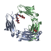
Komagataella pastoris (fungus) / Strain (production host): SMD1168 / References: UniProt: P01860
#2: Polysaccharide beta-D-mannopyranose-(1-4)-2-acetamido-2-deoxy-beta-D-glucopyranose-(1-4)-2-acetamido-2-deoxy-beta- ...beta-D-mannopyranose-(1-4)-2-acetamido-2-deoxy-beta-D-glucopyranose-(1-4)-2-acetamido-2-deoxy-beta-D-glucopyranose


Type: oligosaccharide / Mass: 586.542 Da / Num. of mol.: 2
Source method: isolated from a genetically manipulated source
DescriptorTypeProgram
DManpb1-4DGlcpNAcb1-4DGlcpNAcb1-Glycam Condensed SequenceGMML 1.0
WURCS=2.0/2,3,2/[a2122h-1b_1-5_2*NCC/3=O][a1122h-1b_1-5]/1-1-2/a4-b1_b4-c1WURCSPDB2Glycan 1.1.0
[]{[(4+1)][b-D-GlcpNAc]{[(4+1)][b-D-GlcpNAc]{[(4+1)][b-D-Manp]{}}}}LINUCSPDB-CARE
#3: Chemical ChemComp-FLC / CITRATE ANION / Citric acid


Mass: 189.100 Da / Num. of mol.: 1 / Source method: obtained synthetically / Formula: C6H5O7
#4: Water ChemComp-HOH / water / Water


Mass: 18.015 Da / Num. of mol.: 351 / Source method: isolated from a natural source / Formula: H2O
Sequence detailsThe sequence that was used for this structure comes from the allele IGHG3*11 (accession number ...The sequence that was used for this structure comes from the allele IGHG3*11 (accession number AJ390247). The cDNA of the Fc region of hIgG3 (ATCC: MGC-45809, accession: BC033178) was used for PCR amplification. The Fc sequence belongs to IGHG3*11, *12 allele found in the IMGT database. The expression construct encodes from T225 to K447 (EU numbering). BC033178 also has a Phe residue at position 296.

-
Experimental details

-
Experiment

ExperimentMethod: X-RAY DIFFRACTION / Number of used crystals: 1

-
Sample preparation

CrystalDensity Matthews: 2.6 Å3/Da / Density % sol: 52.75 %
Crystal growTemperature: 293 K / Method: vapor diffusion, sitting drop / pH: 5.5
Details: 20% (w/v) PEG 4000, 100 mM sodium citrate/citric acid, 10% (v/v) 2-propanol

-
Data collection

DiffractionMean temperature: 100 K
Diffraction sourceSource: SYNCHROTRON / Site: APS / Beamline: 17-ID / Wavelength: 1 Å
DetectorType: DECTRIS PILATUS 6M / Detector: PIXEL / Date: Oct 15, 2016
RadiationProtocol: SINGLE WAVELENGTH / Monochromatic (M) / Laue (L): M / Scattering type: x-ray
Radiation wavelengthWavelength: 1 Å / Relative weight: 1
ReflectionResolution: 1.8→37.729 Å / Num. obs: 47374 / % possible obs: 98.4 % / Redundancy: 3.3 % / Biso Wilson estimate: 24.15 Å2 / CC1/2: 0.998 / Rmerge(I) obs: 0.055 / Net I/σ(I): 12.7 / Num. measured all: 156654 / Scaling rejects: 6
Reflection shell
Resolution (Å)Redundancy (%)Rmerge(I) obsCC1/2Diffraction-ID% possible all
1.8-1.843.20.5750.734197.7
9-453.30.0270.999197.8

-
Phasing

PhasingMethod: molecular replacement
Phasing MRModel details: Phaser MODE: MR_AUTO
Highest resolutionLowest resolution
Rotation1.8 Å42.88 Å
Translation1.8 Å42.88 Å

-
Processing

Software
NameVersionClassification
Aimless0.5.27data scaling
PHASER2.6.1phasing
PHENIX1.11_2568refinement
PDB_EXTRACT3.22data extraction
XDSdata reduction
RefinementMethod to determine structure: MOLECULAR REPLACEMENT
Starting model: 4DZ8
Resolution: 1.8→34.729 Å / SU ML: 0.23 / Cross valid method: FREE R-VALUE / σ(F): 1 / Phase error: 22.05
RfactorNum. reflection% reflection
Rfree0.2232 2427 5.13 %
Rwork0.1835 --
obs0.1856 47353 98.3 %
Solvent computationShrinkage radii: 0.9 Å / VDW probe radii: 1.11 Å
Displacement parametersBiso max: 68.67 Å2 / Biso mean: 28.1286 Å2 / Biso min: 10.25 Å2
Refinement stepCycle: final / Resolution: 1.8→34.729 Å
ProteinNucleic acidLigandSolventTotal
Num. atoms3291 0 91 351 3733
Biso mean--41.38 33.37 -
Num. residues----418
Refine LS restraints
Refine-IDTypeDev idealNumber
X-RAY DIFFRACTIONf_bond_d0.0093492
X-RAY DIFFRACTIONf_angle_d1.024773
X-RAY DIFFRACTIONf_chiral_restr0.058548
X-RAY DIFFRACTIONf_plane_restr0.007604
X-RAY DIFFRACTIONf_dihedral_angle_d12.5952103
LS refinement shell

Refine-ID: X-RAY DIFFRACTION / Rfactor Rfree error: 0 / Total num. of bins used: 17

Resolution (Å)Rfactor RfreeNum. reflection RfreeRfactor RworkNum. reflection RworkNum. reflection all% reflection obs (%)
1.8-1.83680.30821200.29342648276898
1.8368-1.87670.31341550.25312625278099
1.8767-1.92030.27191310.23392614274598
1.9203-1.96840.25491460.21262644279099
1.9684-2.02160.25341410.20062638277999
2.0216-2.08110.211530.19392643279699
2.0811-2.14820.22991510.188126372788100
2.1482-2.2250.22361310.1852685281699
2.225-2.3140.24321330.192674280799
2.314-2.41930.22351400.1852634277499
2.4193-2.54690.2481290.17872658278798
2.5469-2.70640.26441520.19262649280199
2.7064-2.91520.24131400.18752666280698
2.9152-3.20840.23911400.19272622276298
3.2084-3.67220.18961710.16722577274896
3.6722-4.62490.19841440.1482620276496
4.6249-34.73530.19681500.18232692284297

+
About Yorodumi

-
News

-
Feb 9, 2022. New format data for meta-information of EMDB entries

New format data for meta-information of EMDB entries

  • Version 3 of the EMDB header file is now the official format.
  • The previous official version 1.9 will be removed from the archive.

Related info.:EMDB header

External links:wwPDB to switch to version 3 of the EMDB data model

-
Aug 12, 2020. Covid-19 info

Covid-19 info

URL: https://pdbj.org/emnavi/covid19.php

New page: Covid-19 featured information page in EM Navigator.

Related info.:Covid-19 info / Mar 5, 2020. Novel coronavirus structure data

+
Mar 5, 2020. Novel coronavirus structure data

Novel coronavirus structure data

Related info.:Yorodumi Speices / Aug 12, 2020. Covid-19 info

External links:COVID-19 featured content - PDBj / Molecule of the Month (242):Coronavirus Proteases

+
Jan 31, 2019. EMDB accession codes are about to change! (news from PDBe EMDB page)

EMDB accession codes are about to change! (news from PDBe EMDB page)

  • The allocation of 4 digits for EMDB accession codes will soon come to an end. Whilst these codes will remain in use, new EMDB accession codes will include an additional digit and will expand incrementally as the available range of codes is exhausted. The current 4-digit format prefixed with “EMD-” (i.e. EMD-XXXX) will advance to a 5-digit format (i.e. EMD-XXXXX), and so on. It is currently estimated that the 4-digit codes will be depleted around Spring 2019, at which point the 5-digit format will come into force.
  • The EM Navigator/Yorodumi systems omit the EMD- prefix.

Related info.:Q: What is EMD? / ID/Accession-code notation in Yorodumi/EM Navigator

External links:EMDB Accession Codes are Changing Soon! / Contact to PDBj

+
Jul 12, 2017. Major update of PDB

Major update of PDB

  • wwPDB released updated PDB data conforming to the new PDBx/mmCIF dictionary.
  • This is a major update changing the version number from 4 to 5, and with Remediation, in which all the entries are updated.
  • In this update, many items about electron microscopy experimental information are reorganized (e.g. em_software).
  • Now, EM Navigator and Yorodumi are based on the updated data.

External links:wwPDB Remediation / Enriched Model Files Conforming to OneDep Data Standards Now Available in the PDB FTP Archive

-
Yorodumi

Thousand views of thousand structures

  • Yorodumi is a browser for structure data from EMDB, PDB, SASBDB, etc.
  • This page is also the successor to EM Navigator detail page, and also detail information page/front-end page for Omokage search.
  • The word "yorodu" (or yorozu) is an old Japanese word meaning "ten thousand". "mi" (miru) is to see.

Related info.:EMDB / PDB / SASBDB / Comparison of 3 databanks / Yorodumi Search / Aug 31, 2016. New EM Navigator & Yorodumi / Yorodumi Papers / Jmol/JSmol / Function and homology information / Changes in new EM Navigator and Yorodumi

Read more