 Movie
Movie Controller

[English] 日本語
 Yorodumi
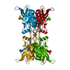
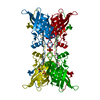
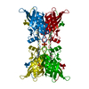
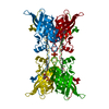
- PDB-5w2k: Crystal structure of mutant CJ YCEI protein (CJ-G34C) with hydrox... -
+ Open data

- Basic information
| Entry | Database: PDB / ID: 5w2k | ||||||
|---|---|---|---|---|---|---|---|
| Title | Crystal structure of mutant CJ YCEI protein (CJ-G34C) with hydroxymercuribenzoic acid guest structure | ||||||
 Components Components | Polyisoprenoid-binding protein | ||||||
 Keywords Keywords | UNKNOWN FUNCTION / nanomaterial nanoporous | ||||||
| Function / homology |  Function and homology information Function and homology informationLipid/polyisoprenoid-binding, YceI-like / Lipid/polyisoprenoid-binding, YceI-like / Lipid/polyisoprenoid-binding, YceI-like superfamily / YceI-like domain / YceI-like domain / Lipocalin / Beta Barrel / Mainly Beta Similarity search - Domain/homology | ||||||
| Biological species |   Campylobacter jejuni (Campylobacter) Campylobacter jejuni (Campylobacter) | ||||||
| Method |  X-RAY DIFFRACTION / SYNCHROTRON / MOLECULAR REPLACEMENT / Resolution: 2.78 Å X-RAY DIFFRACTION / SYNCHROTRON / MOLECULAR REPLACEMENT / Resolution: 2.78 Å | ||||||
 Authors Authors | Huber, T.R. / Snow, C.D. | ||||||
 Citation Citation | Journal: Bioconjug. Chem. / Year: 2018 Title: Installing Guest Molecules at Specific Sites within Scaffold Protein Crystals. Authors: Huber, T.R. / McPherson, E.C. / Keating, C.E. / Snow, C.D. | ||||||
| History |
|
- Structure visualization
| Structure viewer | Molecule: MolmilJmol/JSmol |
|---|
- Downloads & links
-Download
| PDBx/mmCIF format | 5w2k.cif.gz | 50.3 KB | Display | PDBx/mmCIF format |
|---|---|---|---|---|
| PDB format | pdb5w2k.ent.gz | 34 KB | Display | PDB format |
| PDBx/mmJSON format | 5w2k.json.gz | Tree view | PDBx/mmJSON format | |
| Others |  Other downloads Other downloads |
-Validation report
| Summary document | 5w2k_validation.pdf.gz | 454.5 KB | Display | wwPDB validaton report |
|---|---|---|---|---|
| Full document | 5w2k_full_validation.pdf.gz | 455.9 KB | Display | |
| Data in XML | 5w2k_validation.xml.gz | 8.8 KB | Display | |
| Data in CIF | 5w2k_validation.cif.gz | 10.9 KB | Display | |
| Arichive directory | https://data.pdbj.org/pub/pdb/validation_reports/w2/5w2kftp://data.pdbj.org/pub/pdb/validation_reports/w2/5w2k | HTTPS FTP |
-Related structure data
| Related structure data |  5w17C  5w2dSC  5w2rC  5w2vC  5w2xC  5w2zC  5w30C  5w31C  5w32C 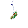 5w37C 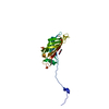 5w39C 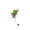 5w3aC 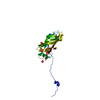 5w3bC 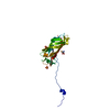 5w3cC C: citing same article ( S: Starting model for refinement |
|---|---|
| Similar structure data |


-Links
 PDBj
PDBj

- Assembly
| Deposited unit | 
| ||||||||
|---|---|---|---|---|---|---|---|---|---|
| 1 | 
| ||||||||
| Unit cell |
|
-Components
| #1: Protein | Mass: 20245.883 Da / Num. of mol.: 1 Source method: isolated from a genetically manipulated source Source: (gene. exp.) Campylobacter jejuni (Campylobacter) / Gene: A0L11_08945, A0M64_02260, A0M70_03345, AD53_02580 / Plasmid: pSB3 / Production host: | ||||
|---|---|---|---|---|---|
| #2: Chemical | ChemComp-UNL / Mass: 282.547 Da / Num. of mol.: 1 / Source method: obtained synthetically | ||||
| #3: Chemical |   Mass: 96.063 Da / Num. of mol.: 2 / Source method: obtained synthetically / Formula: SO4 Mass: 96.063 Da / Num. of mol.: 2 / Source method: obtained synthetically / Formula: SO4#4: Chemical | ChemComp-MBO / | 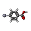  Mass: 321.703 Da / Num. of mol.: 1 / Source method: obtained synthetically / Formula: C7H5HgO2 Mass: 321.703 Da / Num. of mol.: 1 / Source method: obtained synthetically / Formula: C7H5HgO2#5: Water | ChemComp-HOH / |  Mass: 18.015 Da / Num. of mol.: 18 / Source method: isolated from a natural source / Formula: H2O Mass: 18.015 Da / Num. of mol.: 18 / Source method: isolated from a natural source / Formula: H2O |
-Experimental details
-Experiment
| Experiment | Method: X-RAY DIFFRACTION / Number of used crystals: 1 |
|---|
- Sample preparation
| Crystal | Mosaicity: 0.26 ° |
|---|---|
| Crystal grow | Temperature: 293 K / Method: vapor diffusion, sitting drop / pH: 6 / Details: 3.2 M Ammonium Sulfate, 0.1 M Bis-Tris |
-Data collection
| Diffraction | Mean temperature: 100 K | ||||||||||||||||||||||||||||||||||||||||||||||||||||||||||||||||||||||||||||||||||||||||
|---|---|---|---|---|---|---|---|---|---|---|---|---|---|---|---|---|---|---|---|---|---|---|---|---|---|---|---|---|---|---|---|---|---|---|---|---|---|---|---|---|---|---|---|---|---|---|---|---|---|---|---|---|---|---|---|---|---|---|---|---|---|---|---|---|---|---|---|---|---|---|---|---|---|---|---|---|---|---|---|---|---|---|---|---|---|---|---|---|---|
| Diffraction source | Source: SYNCHROTRON / Site: ALS  / Beamline: 4.2.2 / Wavelength: 1 Å / Beamline: 4.2.2 / Wavelength: 1 Å | ||||||||||||||||||||||||||||||||||||||||||||||||||||||||||||||||||||||||||||||||||||||||
| Detector | Type: RDI CMOS_8M / Detector: CMOS / Date: Apr 4, 2017 | ||||||||||||||||||||||||||||||||||||||||||||||||||||||||||||||||||||||||||||||||||||||||
| Radiation | Monochromator: Si (111) double crystal / Protocol: SINGLE WAVELENGTH / Monochromatic (M) / Laue (L): M / Scattering type: x-ray | ||||||||||||||||||||||||||||||||||||||||||||||||||||||||||||||||||||||||||||||||||||||||
| Radiation wavelength | Wavelength: 1 Å / Relative weight: 1 | ||||||||||||||||||||||||||||||||||||||||||||||||||||||||||||||||||||||||||||||||||||||||
| Reflection | Resolution: 2.78→38.714 Å / Num. all: 12465 / Num. obs: 12465 / % possible obs: 99.7 % / Redundancy: 19.6 % / Rpim(I) all: 0.039 / Rrim(I) all: 0.176 / Rsym value: 0.171 / Net I/av σ(I): 3.3 / Net I/σ(I): 11.4 / Num. measured all: 244820 | ||||||||||||||||||||||||||||||||||||||||||||||||||||||||||||||||||||||||||||||||||||||||
| Reflection shell | Diffraction-ID: 1
|
- Processing
| Software |
| |||||||||||||||||||||||||||||||||||||||||||||
|---|---|---|---|---|---|---|---|---|---|---|---|---|---|---|---|---|---|---|---|---|---|---|---|---|---|---|---|---|---|---|---|---|---|---|---|---|---|---|---|---|---|---|---|---|---|---|
| Refinement | Method to determine structure: MOLECULAR REPLACEMENT Starting model: 5W2D Resolution: 2.78→38.71 Å / Cor.coef. Fo:Fc: 0.954 / Cor.coef. Fo:Fc free: 0.935 / Cross valid method: THROUGHOUT / σ(F): 0 / ESU R: 0.296 / ESU R Free: 0.254 / Details: U VALUES : REFINED INDIVIDUALLY
| |||||||||||||||||||||||||||||||||||||||||||||
| Solvent computation | Ion probe radii: 0.8 Å / Shrinkage radii: 0.8 Å / VDW probe radii: 1.2 Å | |||||||||||||||||||||||||||||||||||||||||||||
| Displacement parameters | Biso max: 305.21 Å2 / Biso mean: 92.06 Å2 / Biso min: 55.09 Å2
| |||||||||||||||||||||||||||||||||||||||||||||
| Refinement step | Cycle: final / Resolution: 2.78→38.71 Å
| |||||||||||||||||||||||||||||||||||||||||||||
| Refine LS restraints |
| |||||||||||||||||||||||||||||||||||||||||||||
| LS refinement shell | Resolution: 2.78→2.852 Å / Rfactor Rfree error: 0 / Total num. of bins used: 20
|
