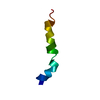
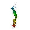
 Movie
Movie Controller

[English] 日本語
 Yorodumi
- PDB-5vxq: X-Ray crystallography structure of the parallel stranded duplex f... -
+ Open data

- Basic information
| Entry | Database: PDB / ID: 5vxq | ||||||||||||||||||||
|---|---|---|---|---|---|---|---|---|---|---|---|---|---|---|---|---|---|---|---|---|---|
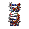
| Title | X-Ray crystallography structure of the parallel stranded duplex formed by 5-rA5-dA-rA5 | ||||||||||||||||||||
 Components Components | DNA/RNA (5'-R(* Keywords KeywordsRNA / DNA / duplex | Function / homology | AMMONIUM ION / DNA/RNA hybrid / DNA/RNA hybrid (> 10) |  Function and homology information Function and homology informationBiological species | synthetic construct (others) | Method |  X-RAY DIFFRACTION / SYNCHROTRON / MOLECULAR REPLACEMENT / Resolution: 1.002 Å X-RAY DIFFRACTION / SYNCHROTRON / MOLECULAR REPLACEMENT / Resolution: 1.002 Å  Authors AuthorsXie, J. / Chen, Y. / Wei, X. / Kozlov, G. / Gehring, K. |  CitationJournal: Nucleic Acids Res. / Year: 2017 CitationJournal: Nucleic Acids Res. / Year: 2017Title: Influence of nucleotide modifications at the C2' position on the Hoogsteen base-paired parallel-stranded duplex of poly(A) RNA. Authors: Copp, W. / Denisov, A.Y. / Xie, J. / Noronha, A.M. / Liczner, C. / Safaee, N. / Wilds, C.J. / Gehring, K. History |
|
- Structure visualization
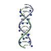
| Structure viewer | Molecule: MolmilJmol/JSmol |
|---|
- Downloads & links
-Download
| PDBx/mmCIF format | 5vxq.cif.gz | 37.3 KB | Display | PDBx/mmCIF format |
|---|---|---|---|---|
| PDB format | pdb5vxq.ent.gz | 27.6 KB | Display | PDB format |
| PDBx/mmJSON format | 5vxq.json.gz | Tree view | PDBx/mmJSON format | |
| Others |  Other downloads Other downloads |
-Validation report
| Arichive directory | https://data.pdbj.org/pub/pdb/validation_reports/vx/5vxqftp://data.pdbj.org/pub/pdb/validation_reports/vx/5vxq | HTTPS FTP |
|---|
-Related structure data


-Links
 PDBj
PDBj


- Assembly
| Deposited unit | 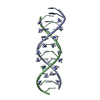
| ||||||||
|---|---|---|---|---|---|---|---|---|---|
| 1 |
| ||||||||
| Unit cell |
| ||||||||
| Components on special symmetry positions |
|
-Components
| #1: DNA/RNA hybrid | Mass: 3560.307 Da / Num. of mol.: 2 / Source method: obtained synthetically / Details: modified RNA duplex / Source: (synth.) synthetic construct (others) #2: Chemical | ChemComp-NH4 /   Mass: 18.038 Da / Num. of mol.: 18 / Source method: obtained synthetically / Formula: H4N Mass: 18.038 Da / Num. of mol.: 18 / Source method: obtained synthetically / Formula: H4N#3: Water | ChemComp-HOH / |  Mass: 18.015 Da / Num. of mol.: 71 / Source method: isolated from a natural source / Formula: H2O Mass: 18.015 Da / Num. of mol.: 71 / Source method: isolated from a natural source / Formula: H2O |
|---|
-Experimental details
-Experiment
| Experiment | Method: X-RAY DIFFRACTION / Number of used crystals: 1 |
|---|
- Sample preparation
| Crystal | Density Matthews: 1.5 Å3/Da / Density % sol: 17.84 % |
|---|---|
| Crystal grow | Temperature: 295 K / Method: hanging drop, vapor diffusion / pH: 5 Details: 0.5 uL 5-rA5-dA-rA5 and RRM23 (98-269) in 10 mM HEPES, 100 mM sodium chloride, 2 mM DTT, pH 7.0 + 0.5 uL 1.6 M ammonium sulfate, 0.1 M citric acid, pH 5.0 |
-Data collection
| Diffraction | Mean temperature: 77 K |
|---|---|
| Diffraction source | Source: SYNCHROTRON / Site: CLSI  / Beamline: 08ID-1 / Wavelength: 1.28206 Å / Beamline: 08ID-1 / Wavelength: 1.28206 Å |
| Detector | Type: RAYONIX MX-300 / Detector: CCD / Date: May 1, 2015 |
| Radiation | Monochromator: double crystal Si(111) / Protocol: SINGLE WAVELENGTH / Monochromatic (M) / Laue (L): M / Scattering type: x-ray |
| Radiation wavelength | Wavelength: 1.28206 Å / Relative weight: 1 |
| Reflection | Resolution: 1.002→22.58 Å / Num. obs: 23843 / % possible obs: 96.4 % / Redundancy: 1 % / Net I/σ(I): 17.65 |
| Reflection shell | Highest resolution: 1.002 Å |
- Processing
| Software |
| ||||||||||||||||
|---|---|---|---|---|---|---|---|---|---|---|---|---|---|---|---|---|---|
| Refinement | Method to determine structure: MOLECULAR REPLACEMENT / Resolution: 1.002→22.58 Å / Cross valid method: FREE R-VALUE
| ||||||||||||||||
| Refinement step | Cycle: LAST / Resolution: 1.002→22.58 Å
|
