 Movie
Movie Controller

[English] 日本語
 Yorodumi
- PDB-5vvv: Structural Investigations of the Substrate Specificity of Human O... -
+ Open data

- Basic information
| Entry | Database: PDB / ID: 5vvv | ||||||
|---|---|---|---|---|---|---|---|
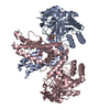
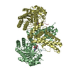
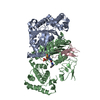
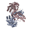
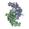
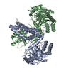
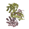
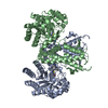
| Title | Structural Investigations of the Substrate Specificity of Human O-GlcNAcase | ||||||
 Components Components |
| ||||||
 Keywords Keywords | HYDROLASE / OGA / Human O-GlcNAcase / a-crystalline B | ||||||
| Function / homology |  Function and homology information Function and homology informationmicrotubule polymerization or depolymerization / negative regulation of intracellular transport / hyalurononglucosaminidase activity / glycoprotein metabolic process / hexosaminidase activity / N-acetylglucosamine metabolic process / apoptotic process involved in morphogenesis / protein deglycosylation / protein O-GlcNAcase / [protein]-3-O-(N-acetyl-D-glucosaminyl)-L-serine/L-threonine O-N-acetyl-alpha-D-glucosaminase activity ...microtubule polymerization or depolymerization / negative regulation of intracellular transport / hyalurononglucosaminidase activity / glycoprotein metabolic process / hexosaminidase activity / N-acetylglucosamine metabolic process / apoptotic process involved in morphogenesis / protein deglycosylation / protein O-GlcNAcase / [protein]-3-O-(N-acetyl-D-glucosaminyl)-L-serine/L-threonine O-N-acetyl-alpha-D-glucosaminase activity / regulation of programmed cell death / glycoprotein catabolic process / cardiac myofibril / structural constituent of eye lens / tubulin complex assembly / protein O-linked glycosylation / negative regulation of amyloid fibril formation / lens development in camera-type eye / M band / muscle organ development / actin filament bundle / negative regulation of reactive oxygen species metabolic process / HSF1-dependent transactivation / stress-activated MAPK cascade / negative regulation of protein-containing complex assembly / muscle contraction / synaptic membrane / glutathione metabolic process / response to hydrogen peroxide / cellular response to gamma radiation / negative regulation of cell growth / Z disc / : / response to estradiol / amyloid-beta binding / response to heat / protein refolding / protein folding / microtubule binding / dendritic spine / perikaryon / response to hypoxia / lysosome / protein stabilization / negative regulation of gene expression / axon / negative regulation of DNA-templated transcription / negative regulation of apoptotic process / protein-containing complex binding / structural molecule activity / cell surface / protein homodimerization activity / protein-containing complex / mitochondrion / extracellular exosome / nucleoplasm / membrane / metal ion binding / identical protein binding / nucleus / cytoplasm / cytosol Similarity search - Function | ||||||
| Biological species |  Homo sapiens (human) Homo sapiens (human) | ||||||
| Method |  X-RAY DIFFRACTION / SYNCHROTRON / MOLECULAR REPLACEMENT / Resolution: 2.8 Å X-RAY DIFFRACTION / SYNCHROTRON / MOLECULAR REPLACEMENT / Resolution: 2.8 Å | ||||||
 Authors Authors | Li, B. / Jiang, J. / Li, H. / Hu, C.-W. | ||||||
| Funding support |  United States, 1items United States, 1items
| ||||||
 Citation Citation | Journal: Nat Commun / Year: 2017 Title: Structural insights into the substrate binding adaptability and specificity of human O-GlcNAcase. Authors: Li, B. / Li, H. / Hu, C.W. / Jiang, J. | ||||||
| History |
|
- Structure visualization
| Structure viewer | Molecule: MolmilJmol/JSmol |
|---|
- Downloads & links
-Download
| PDBx/mmCIF format | 5vvv.cif.gz | 188.2 KB | Display | PDBx/mmCIF format |
|---|---|---|---|---|
| PDB format | pdb5vvv.ent.gz | 146.7 KB | Display | PDB format |
| PDBx/mmJSON format | 5vvv.json.gz | Tree view | PDBx/mmJSON format | |
| Others |  Other downloads Other downloads |
-Validation report
| Arichive directory | https://data.pdbj.org/pub/pdb/validation_reports/vv/5vvvftp://data.pdbj.org/pub/pdb/validation_reports/vv/5vvv | HTTPS FTP |
|---|
-Related structure data
| Related structure data | 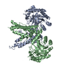 5vvoC 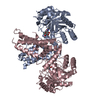 5vvtC  5vvuC  5vvxC  5un8S C: citing same article ( S: Starting model for refinement |
|---|---|
| Similar structure data |


-Links
 PDBj
PDBj


- Assembly
| Deposited unit | 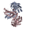
| ||||||||||||||||||
|---|---|---|---|---|---|---|---|---|---|---|---|---|---|---|---|---|---|---|---|
| 1 |
| ||||||||||||||||||
| Unit cell |
| ||||||||||||||||||
| Noncrystallographic symmetry (NCS) | NCS domain:
NCS domain segments: Component-ID: _ / Ens-ID: 1 / Beg auth comp-ID: HIS / Beg label comp-ID: HIS / End auth comp-ID: PRO / End label comp-ID: PRO / Refine code: _ / Auth seq-ID: 59 - 694 / Label seq-ID: 1 - 494
|
-Components
| #1: Protein | Mass: 57822.582 Da / Num. of mol.: 2 / Fragment: UNP residues 60-400, 553-704 Source method: isolated from a genetically manipulated source Source: (gene. exp.) Homo sapiens (human) / Gene: MGEA5, HEXC, KIAA0679, MEA5 / Production host:  References: UniProt: O60502, protein O-GlcNAcase, Hydrolases; Glycosylases; Glycosidases, i.e. enzymes that hydrolyse O- and S-glycosyl compounds #2: Protein/peptide | Mass: 1516.716 Da / Num. of mol.: 2 / Source method: obtained synthetically / Source: (synth.) Homo sapiens (human) / References: UniProt: P02511*PLUS#3: Sugar |   Type: D-saccharide, beta linking / Mass: 221.208 Da / Num. of mol.: 2 / Source method: obtained synthetically / Formula: C8H15NO6 Type: D-saccharide, beta linking / Mass: 221.208 Da / Num. of mol.: 2 / Source method: obtained synthetically / Formula: C8H15NO6#4: Water | ChemComp-HOH / |  Mass: 18.015 Da / Num. of mol.: 35 / Source method: isolated from a natural source / Formula: H2O Mass: 18.015 Da / Num. of mol.: 35 / Source method: isolated from a natural source / Formula: H2OHas protein modification | Y | |
|---|
-Experimental details
-Experiment
| Experiment | Method: X-RAY DIFFRACTION / Number of used crystals: 1 |
|---|
- Sample preparation
| Crystal | Density Matthews: 2.76 Å3/Da / Density % sol: 55.4 % |
|---|---|
| Crystal grow | Temperature: 293 K / Method: evaporation Details: 0.024 M ammonium citrate tribasic (pH 7.0), 0.015 M MES monohydrate, 0.096 M potassium thiocyanate, 0.25 M Sodium acetate trihydrate, 0.037 M imidazole, 0.002 M zinc sulfate heptahydrate, 9. ...Details: 0.024 M ammonium citrate tribasic (pH 7.0), 0.015 M MES monohydrate, 0.096 M potassium thiocyanate, 0.25 M Sodium acetate trihydrate, 0.037 M imidazole, 0.002 M zinc sulfate heptahydrate, 9.6 % w/v polyethylene glycol 3,350, 2.4 % w/v polyethylene glycol monomethyl ether 2,000, and 4% w/v polyethylene glycol monomethyl ether 550 |
-Data collection
| Diffraction | Mean temperature: 80 K |
|---|---|
| Diffraction source | Source: SYNCHROTRON / Site: APS / Beamline: 21-ID-G / Wavelength: 0.978 Å |
| Detector | Type: MARMOSAIC 300 mm CCD / Detector: CCD / Date: Feb 22, 2017 |
| Radiation | Protocol: SINGLE WAVELENGTH / Monochromatic (M) / Laue (L): M / Scattering type: x-ray |
| Radiation wavelength | Wavelength: 0.978 Å / Relative weight: 1 |
| Reflection | Resolution: 2.7→50 Å / Num. obs: 31917 / % possible obs: 99.8 % / Redundancy: 5.6 % / Rpim(I) all: 0.049 / Net I/σ(I): 1.5 |
| Reflection shell | Resolution: 2.7→2.8 Å / Redundancy: 5.5 % / Num. unique obs: 1705 / CC1/2: 0.58 / Rpim(I) all: 0.341 / % possible all: 98.6 |
- Processing
| Software |
| ||||||||||||||||||||||||||||||||||||||||||||||||||||||||||||||||||||||||||||||||||||||||||||||||||||||||||||||||||||||||||||||||||||||||||||||||||||||||||||||||||||||||||||||||||||||
|---|---|---|---|---|---|---|---|---|---|---|---|---|---|---|---|---|---|---|---|---|---|---|---|---|---|---|---|---|---|---|---|---|---|---|---|---|---|---|---|---|---|---|---|---|---|---|---|---|---|---|---|---|---|---|---|---|---|---|---|---|---|---|---|---|---|---|---|---|---|---|---|---|---|---|---|---|---|---|---|---|---|---|---|---|---|---|---|---|---|---|---|---|---|---|---|---|---|---|---|---|---|---|---|---|---|---|---|---|---|---|---|---|---|---|---|---|---|---|---|---|---|---|---|---|---|---|---|---|---|---|---|---|---|---|---|---|---|---|---|---|---|---|---|---|---|---|---|---|---|---|---|---|---|---|---|---|---|---|---|---|---|---|---|---|---|---|---|---|---|---|---|---|---|---|---|---|---|---|---|---|---|---|---|
| Refinement | Method to determine structure: MOLECULAR REPLACEMENT Starting model: 5UN8 Resolution: 2.8→50 Å / Cor.coef. Fo:Fc: 0.946 / Cor.coef. Fo:Fc free: 0.903 / SU B: 16.196 / SU ML: 0.306 / Cross valid method: THROUGHOUT / ESU R: 0.984 / ESU R Free: 0.352
| ||||||||||||||||||||||||||||||||||||||||||||||||||||||||||||||||||||||||||||||||||||||||||||||||||||||||||||||||||||||||||||||||||||||||||||||||||||||||||||||||||||||||||||||||||||||
| Solvent computation | Ion probe radii: 0.8 Å / Shrinkage radii: 0.8 Å / VDW probe radii: 1.2 Å | ||||||||||||||||||||||||||||||||||||||||||||||||||||||||||||||||||||||||||||||||||||||||||||||||||||||||||||||||||||||||||||||||||||||||||||||||||||||||||||||||||||||||||||||||||||||
| Displacement parameters | Biso mean: 74.527 Å2
| ||||||||||||||||||||||||||||||||||||||||||||||||||||||||||||||||||||||||||||||||||||||||||||||||||||||||||||||||||||||||||||||||||||||||||||||||||||||||||||||||||||||||||||||||||||||
| Refinement step | Cycle: 1 / Resolution: 2.8→50 Å
| ||||||||||||||||||||||||||||||||||||||||||||||||||||||||||||||||||||||||||||||||||||||||||||||||||||||||||||||||||||||||||||||||||||||||||||||||||||||||||||||||||||||||||||||||||||||
| Refine LS restraints |
|