 Movie
Movie Controller

[English] 日本語
 Yorodumi
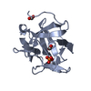
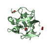
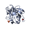
- PDB-5vbk: Crystal structure of a galactose-binding Lectin from Mytilus cali... -
+ Open data

- Basic information
| Entry | Database: PDB / ID: 5vbk | ||||||
|---|---|---|---|---|---|---|---|
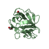
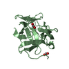
| Title | Crystal structure of a galactose-binding Lectin from Mytilus californianus | ||||||
 Components Components | Lectin | ||||||
 Keywords Keywords | SUGAR BINDING PROTEIN / Galactose-Binding Lectin | ||||||
| Function / homology | galactose binding / Trefoil (Acidic Fibroblast Growth Factor, subunit A) - #50 / Trefoil (Acidic Fibroblast Growth Factor, subunit A) / Trefoil / Mainly Beta / Galactose-binding lectin Function and homology information Function and homology information | ||||||
| Biological species |  Mytilus californianus (California mussel) Mytilus californianus (California mussel) | ||||||
| Method |  X-RAY DIFFRACTION / MOLECULAR REPLACEMENT / Resolution: 1.788 Å X-RAY DIFFRACTION / MOLECULAR REPLACEMENT / Resolution: 1.788 Å | ||||||
 Authors Authors | Hernandez-Santoyo, A. / Garcia-Maldonado, E. | ||||||
 Citation Citation | Journal: Fish Shellfish Immunol. / Year: 2017 Title: Molecular and functional characterization of a glycosylated Galactose-Binding lectin from Mytilus californianus. Authors: Garcia-Maldonado, E. / Cano-Sanchez, P. / Hernandez-Santoyo, A. | ||||||
| History |
|
- Structure visualization
| Structure viewer | Molecule: MolmilJmol/JSmol |
|---|
- Downloads & links
-Download
| PDBx/mmCIF format | 5vbk.cif.gz | 78.5 KB | Display | PDBx/mmCIF format |
|---|---|---|---|---|
| PDB format | pdb5vbk.ent.gz | 58.1 KB | Display | PDB format |
| PDBx/mmJSON format | 5vbk.json.gz | Tree view | PDBx/mmJSON format | |
| Others |  Other downloads Other downloads |
-Validation report
| Summary document | 5vbk_validation.pdf.gz | 439.8 KB | Display | wwPDB validaton report |
|---|---|---|---|---|
| Full document | 5vbk_full_validation.pdf.gz | 441.4 KB | Display | |
| Data in XML | 5vbk_validation.xml.gz | 15.4 KB | Display | |
| Data in CIF | 5vbk_validation.cif.gz | 21.8 KB | Display | |
| Arichive directory | https://data.pdbj.org/pub/pdb/validation_reports/vb/5vbkftp://data.pdbj.org/pub/pdb/validation_reports/vb/5vbk | HTTPS FTP |
-Related structure data
| Related structure data |  3wmvS S: Starting model for refinement |
|---|---|
| Similar structure data |


-Links
 PDBj
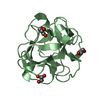
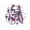
PDBj- Assembly
| Deposited unit | 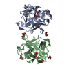
| ||||||||
|---|---|---|---|---|---|---|---|---|---|
| 1 | 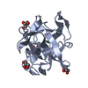
| ||||||||
| 2 | 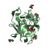
| ||||||||
| Unit cell |
|
-Components
| #1: Protein | Mass: 16943.363 Da / Num. of mol.: 2 / Source method: isolated from a natural source / Source: (natural) Mytilus californianus (California mussel) / References: UniProt: A0A0P0E482#2: Chemical | ChemComp-GOL /   Mass: 92.094 Da / Num. of mol.: 9 / Source method: obtained synthetically / Formula: C3H8O3 Mass: 92.094 Da / Num. of mol.: 9 / Source method: obtained synthetically / Formula: C3H8O3#3: Water | ChemComp-HOH / |  Mass: 18.015 Da / Num. of mol.: 207 / Source method: isolated from a natural source / Formula: H2O Mass: 18.015 Da / Num. of mol.: 207 / Source method: isolated from a natural source / Formula: H2O |
|---|
-Experimental details
-Experiment
| Experiment | Method: X-RAY DIFFRACTION / Number of used crystals: 1 |
|---|
- Sample preparation
| Crystal | Density Matthews: 2.19 Å3/Da / Density % sol: 43.73 % |
|---|---|
| Crystal grow | Temperature: 291 K / Method: vapor diffusion, sitting drop Details: 0.2M potassium thiocyanate, 0.1 M Bis-Tris Propane pH 8.5, 20% PEG 3350 |
-Data collection
| Diffraction | Mean temperature: 101 K |
|---|---|
| Diffraction source | Source: ROTATING ANODE / Type: RIGAKU MICROMAX-007 / Wavelength: 1.54 Å |
| Detector | Type: DECTRIS PILATUS 200K / Detector: PIXEL / Date: Dec 1, 2016 |
| Radiation | Protocol: SINGLE WAVELENGTH / Monochromatic (M) / Laue (L): M / Scattering type: x-ray |
| Radiation wavelength | Wavelength: 1.54 Å / Relative weight: 1 |
| Reflection | Resolution: 1.78→50 Å / Num. obs: 26332 / % possible obs: 95.06 % / Redundancy: 4.4 % / CC1/2: 0.85 / Rmerge(I) obs: 0.06 / Rpim(I) all: 0.04 / Net I/σ(I): 28.8 |
- Processing
| Software |
| ||||||||||||||||||||||||||||||||||||||||||||||||||||||||||||||||||||||
|---|---|---|---|---|---|---|---|---|---|---|---|---|---|---|---|---|---|---|---|---|---|---|---|---|---|---|---|---|---|---|---|---|---|---|---|---|---|---|---|---|---|---|---|---|---|---|---|---|---|---|---|---|---|---|---|---|---|---|---|---|---|---|---|---|---|---|---|---|---|---|---|
| Refinement | Method to determine structure: MOLECULAR REPLACEMENT Starting model: 3WMV Resolution: 1.788→35.088 Å / SU ML: 0.22 / Cross valid method: FREE R-VALUE / σ(F): 1.34 / Phase error: 25.36
| ||||||||||||||||||||||||||||||||||||||||||||||||||||||||||||||||||||||
| Solvent computation | Shrinkage radii: 0.9 Å / VDW probe radii: 1.11 Å | ||||||||||||||||||||||||||||||||||||||||||||||||||||||||||||||||||||||
| Refinement step | Cycle: LAST / Resolution: 1.788→35.088 Å
| ||||||||||||||||||||||||||||||||||||||||||||||||||||||||||||||||||||||
| Refine LS restraints |
| ||||||||||||||||||||||||||||||||||||||||||||||||||||||||||||||||||||||
| LS refinement shell |
|
