 Movie
Movie Controller

[English] 日本語
 Yorodumi
- PDB-5v7p: Atomic structure of the eukaryotic intramembrane Ras methyltransf... -
+ Open data

- Basic information
| Entry | Database: PDB / ID: 5v7p | ||||||
|---|---|---|---|---|---|---|---|
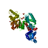
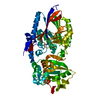
| Title | Atomic structure of the eukaryotic intramembrane Ras methyltransferase ICMT (isoprenylcysteine carboxyl methyltransferase), in complex with a monobody | ||||||
 Components Components |
| ||||||
 Keywords Keywords | TRANSFERASE / membrane protein / membrane enzyme / methyltransferase / LCP | ||||||
| Function / homology |  Function and homology information Function and homology informationprotein-S-isoprenylcysteine O-methyltransferase / protein C-terminal S-isoprenylcysteine carboxyl O-methyltransferase activity / methylation / endoplasmic reticulum membrane / endoplasmic reticulum Similarity search - Function | ||||||
| Biological species |  synthetic construct (others) | ||||||
| Method |  X-RAY DIFFRACTION / SYNCHROTRON / SIRAS / Resolution: 2.3 Å X-RAY DIFFRACTION / SYNCHROTRON / SIRAS / Resolution: 2.3 Å | ||||||
 Authors Authors | Long, S.B. / Diver, M.M. / Pedi, L. / Koide, A. / Koide, S. | ||||||
 Citation Citation | Journal: Nature / Year: 2018 Title: Atomic structure of the eukaryotic intramembrane RAS methyltransferase ICMT. Authors: Diver, M.M. / Pedi, L. / Koide, A. / Koide, S. / Long, S.B. | ||||||
| History |
|
- Structure visualization
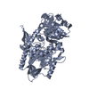
| Structure viewer | Molecule: MolmilJmol/JSmol |
|---|
- Downloads & links
-Download
| PDBx/mmCIF format | 5v7p.cif.gz | 100.3 KB | Display | PDBx/mmCIF format |
|---|---|---|---|---|
| PDB format | pdb5v7p.ent.gz | 76.7 KB | Display | PDB format |
| PDBx/mmJSON format | 5v7p.json.gz | Tree view | PDBx/mmJSON format | |
| Others |  Other downloads Other downloads |
-Validation report
| Arichive directory | https://data.pdbj.org/pub/pdb/validation_reports/v7/5v7pftp://data.pdbj.org/pub/pdb/validation_reports/v7/5v7p | HTTPS FTP |
|---|
-Related structure data


-Links
 PDBj
PDBj

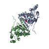
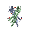
- Assembly
| Deposited unit | 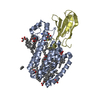
| ||||||||
|---|---|---|---|---|---|---|---|---|---|
| 1 |
| ||||||||
| Unit cell |
|
-Components
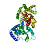
-Protein , 2 types, 2 molecules AD
| #1: Protein | Mass: 32857.156 Da / Num. of mol.: 1 Source method: isolated from a genetically manipulated source Source: (gene. exp.)  Pichia (fungus) Pichia (fungus)References: UniProt: D6WJ77, protein-S-isoprenylcysteine O-methyltransferase |
|---|---|
| #2: Protein | Mass: 10135.358 Da / Num. of mol.: 1 Source method: isolated from a genetically manipulated source Source: (gene. exp.) synthetic construct (others) / Production host:  |
-Non-polymers , 5 types, 117 molecules 
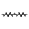


| #3: Chemical | ChemComp-SAH /  Type: L-peptide linking / Mass: 384.411 Da / Num. of mol.: 1 / Source method: obtained synthetically / Formula: C14H20N6O5S Type: L-peptide linking / Mass: 384.411 Da / Num. of mol.: 1 / Source method: obtained synthetically / Formula: C14H20N6O5S | ||||||
|---|---|---|---|---|---|---|---|
| #4: Chemical |  Mass: 156.308 Da / Num. of mol.: 3 / Source method: obtained synthetically / Formula: C11H24 Mass: 156.308 Da / Num. of mol.: 3 / Source method: obtained synthetically / Formula: C11H24#5: Chemical | ChemComp-D10 /  Mass: 142.282 Da / Num. of mol.: 11 / Source method: obtained synthetically / Formula: C10H22 Mass: 142.282 Da / Num. of mol.: 11 / Source method: obtained synthetically / Formula: C10H22#6: Chemical | ChemComp-MPG / [(  Mass: 356.540 Da / Num. of mol.: 10 / Source method: obtained synthetically / Formula: C21H40O4 Mass: 356.540 Da / Num. of mol.: 10 / Source method: obtained synthetically / Formula: C21H40O4#7: Water | ChemComp-HOH / | Mass: 18.015 Da / Num. of mol.: 92 / Source method: isolated from a natural source / Formula: H2O |
-Experimental details
-Experiment
| Experiment | Method: X-RAY DIFFRACTION / Number of used crystals: 1 |
|---|
- Sample preparation
| Crystal | Density Matthews: 3.05 Å3/Da / Density % sol: 59.72 % |
|---|---|
| Crystal grow | Temperature: 293 K / Method: lipidic cubic phase Details: 40/60% (v/v ICMT:9.9 monoacylgylcerol) 30% PEG 400 100 mM NaCl 100 mM Na HEPES, pH 7.5 |
-Data collection
| Diffraction | Mean temperature: 90 K |
|---|---|
| Diffraction source | Source: SYNCHROTRON / Site: APS  / Beamline: 24-ID-C / Wavelength: 0.979 Å / Beamline: 24-ID-C / Wavelength: 0.979 Å |
| Detector | Type: DECTRIS PILATUS 6M-F / Detector: PIXEL / Date: Nov 9, 2015 |
| Radiation | Monochromator: Si / Protocol: SINGLE WAVELENGTH / Monochromatic (M) / Laue (L): M / Scattering type: x-ray |
| Radiation wavelength | Wavelength: 0.979 Å / Relative weight: 1 |
| Reflection | Resolution: 2.3→31 Å / Num. obs: 23204 / % possible obs: 97.2 % / Redundancy: 11.2 % / Biso Wilson estimate: 38 Å2 / CC1/2: 0.863 / Rmerge(I) obs: 0.2 / Rpim(I) all: 0.062 / Χ2: 1.01 / Net I/σ(I): 13.3 |
| Reflection shell | Resolution: 2.3→2.35 Å / Redundancy: 11.1 % / Rmerge(I) obs: 0.979 / Mean I/σ(I) obs: 2.5 / Num. unique obs: 1197 / CC1/2: 0.547 / Rpim(I) all: 0.295 / Χ2: 0.95 / % possible all: 99.8 |
- Processing
| Software |
| |||||||||||||||||||||||||||||||||||||||||||||||||||||||||||||||
|---|---|---|---|---|---|---|---|---|---|---|---|---|---|---|---|---|---|---|---|---|---|---|---|---|---|---|---|---|---|---|---|---|---|---|---|---|---|---|---|---|---|---|---|---|---|---|---|---|---|---|---|---|---|---|---|---|---|---|---|---|---|---|---|---|
| Refinement | Method to determine structure: SIRAS / Resolution: 2.3→29.772 Å / SU ML: 0.29 / Cross valid method: FREE R-VALUE / σ(F): 0.3 / Phase error: 22.02 / Stereochemistry target values: ML
| |||||||||||||||||||||||||||||||||||||||||||||||||||||||||||||||
| Solvent computation | Shrinkage radii: 0.9 Å / VDW probe radii: 1.11 Å / Solvent model: FLAT BULK SOLVENT MODEL | |||||||||||||||||||||||||||||||||||||||||||||||||||||||||||||||
| Refinement step | Cycle: LAST / Resolution: 2.3→29.772 Å
| |||||||||||||||||||||||||||||||||||||||||||||||||||||||||||||||
| Refine LS restraints |
| |||||||||||||||||||||||||||||||||||||||||||||||||||||||||||||||
| LS refinement shell |
|
