 Movie
Movie Controller

[English] 日本語
 Yorodumi
- PDB-5v0m: SeMet crystal structure of the Neisseria meningitidis non-core mi... -
+ Open data

- Basic information
| Entry | Database: PDB / ID: 5v0m | ||||||
|---|---|---|---|---|---|---|---|
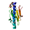
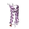
| Title | SeMet crystal structure of the Neisseria meningitidis non-core minor pilin PilV in the monoclinic form | ||||||
 Components Components | Type IV pilin protein | ||||||
 Keywords Keywords | CELL ADHESION / pilin / minor pilin / type IV pili / Neisseria meningitidis | ||||||
| Function / homology | Type IV pilus non-core minor pilin PilE-like / Type IV minor pilin ComP, DNA uptake sequence receptor / type IV pilus assembly / Prokaryotic N-terminal methylation site. / Prokaryotic N-terminal methylation motif / Prokaryotic N-terminal methylation site / Pilin-like / Type IV pilin protein Function and homology information Function and homology information | ||||||
| Biological species |  Neisseria meningitidis (bacteria) Neisseria meningitidis (bacteria) | ||||||
| Method |  X-RAY DIFFRACTION / SYNCHROTRON / MOLECULAR REPLACEMENT / Resolution: 1.407 Å X-RAY DIFFRACTION / SYNCHROTRON / MOLECULAR REPLACEMENT / Resolution: 1.407 Å | ||||||
 Authors Authors | Kolappan, S. / Craig, L. | ||||||
| Funding support |  Canada, 1items Canada, 1items
| ||||||
 Citation Citation | Journal: To Be Published Title: Crystal structure of PilV from Neisseria meningitidis Authors: Kolappan, S. / Craig, L. | ||||||
| History |
|
- Structure visualization
| Structure viewer | Molecule: MolmilJmol/JSmol |
|---|
- Downloads & links
-Download
| PDBx/mmCIF format | 5v0m.cif.gz | 54.9 KB | Display | PDBx/mmCIF format |
|---|---|---|---|---|
| PDB format | pdb5v0m.ent.gz | 39.7 KB | Display | PDB format |
| PDBx/mmJSON format | 5v0m.json.gz | Tree view | PDBx/mmJSON format | |
| Others |  Other downloads Other downloads |
-Validation report
| Arichive directory | https://data.pdbj.org/pub/pdb/validation_reports/v0/5v0mftp://data.pdbj.org/pub/pdb/validation_reports/v0/5v0m | HTTPS FTP |
|---|
-Related structure data
| Related structure data |  5v23S S: Starting model for refinement |
|---|---|
| Similar structure data |

-Links
 PDBj
PDBj
- Assembly
| Deposited unit | 
| ||||||||
|---|---|---|---|---|---|---|---|---|---|
| 1 |
| ||||||||
| Unit cell |
|
-Components
| #1: Protein | Mass: 11124.064 Da / Num. of mol.: 1 Source method: isolated from a genetically manipulated source Source: (gene. exp.) Neisseria meningitidis (bacteria) / Gene: A6L27_01160 / Production host: |
|---|---|
| #2: Chemical | ChemComp-GOL /   Mass: 92.094 Da / Num. of mol.: 1 / Source method: obtained synthetically / Formula: C3H8O3 Mass: 92.094 Da / Num. of mol.: 1 / Source method: obtained synthetically / Formula: C3H8O3 |
| #3: Water | ChemComp-HOH /  Mass: 18.015 Da / Num. of mol.: 106 / Source method: isolated from a natural source / Formula: H2O Mass: 18.015 Da / Num. of mol.: 106 / Source method: isolated from a natural source / Formula: H2O |
| Has protein modification | Y |
-Experimental details
-Experiment
| Experiment | Method: X-RAY DIFFRACTION / Number of used crystals: 1 |
|---|
- Sample preparation
| Crystal | Density Matthews: 1.75 Å3/Da / Density % sol: 33.3 % |
|---|---|
| Crystal grow | Temperature: 293 K / Method: vapor diffusion, hanging drop / pH: 5 Details: 100 mM Sodium Acetate pH 5, 100 mM ammonium chloride, 28 % w/v PEG 8000 |
-Data collection
| Diffraction | Mean temperature: 100 K |
|---|---|
| Diffraction source | Source: SYNCHROTRON / Site: SSRL  / Beamline: BL12-2 / Wavelength: 0.96859 Å / Beamline: BL12-2 / Wavelength: 0.96859 Å |
| Detector | Type: DECTRIS PILATUS 6M / Detector: PIXEL / Date: Feb 2, 2017 |
| Radiation | Protocol: SINGLE WAVELENGTH / Monochromatic (M) / Laue (L): M / Scattering type: x-ray |
| Radiation wavelength | Wavelength: 0.96859 Å / Relative weight: 1 |
| Reflection | Resolution: 1.407→40.63 Å / Num. obs: 15424 / % possible obs: 95.9 % / Redundancy: 5.9 % / CC1/2: 0.997 / Rmerge(I) obs: 0.098 / Rpim(I) all: 0.043 / Net I/σ(I): 12.2 |
| Reflection shell | Resolution: 1.41→1.43 Å / Redundancy: 6 % / Rmerge(I) obs: 0.819 / Mean I/σ(I) obs: 4 / Num. unique all: 772 / CC1/2: 0.843 / Rpim(I) all: 0.359 / % possible all: 97 |
- Processing
| Software |
| |||||||||||||||||||||||||||||||||||||||||||||||||
|---|---|---|---|---|---|---|---|---|---|---|---|---|---|---|---|---|---|---|---|---|---|---|---|---|---|---|---|---|---|---|---|---|---|---|---|---|---|---|---|---|---|---|---|---|---|---|---|---|---|---|
| Refinement | Method to determine structure: MOLECULAR REPLACEMENT Starting model: 5V23 Resolution: 1.407→27.084 Å / SU ML: 0.14 / Cross valid method: FREE R-VALUE / σ(F): 1.33 / Phase error: 22.61
| |||||||||||||||||||||||||||||||||||||||||||||||||
| Solvent computation | Shrinkage radii: 0.9 Å / VDW probe radii: 1.11 Å | |||||||||||||||||||||||||||||||||||||||||||||||||
| Refinement step | Cycle: LAST / Resolution: 1.407→27.084 Å
| |||||||||||||||||||||||||||||||||||||||||||||||||
| Refine LS restraints |
| |||||||||||||||||||||||||||||||||||||||||||||||||
| LS refinement shell |
|
