[English] 日本語
Yorodumi
- PDB-5uyz: Structure of Human T-complex protein 1 subunit epsilon (CCT5) mut... -

+
Open data


ID or keywords:

Loading...

-
Basic information

Entry
Database: PDB / ID: 5uyz
Title
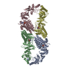
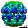
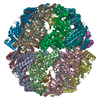
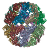
Structure of Human T-complex protein 1 subunit epsilon (CCT5) mutant His147Arg
ComponentsT-complex protein 1 subunit epsilon
KeywordsPROTEIN BINDING / chaperonin Hexadecameric complex ATP-dependent CCT5 gene
Function / homology
Function and homology information


positive regulation of establishment of protein localization to telomere / positive regulation of protein localization to Cajal body / BBSome-mediated cargo-targeting to cilium / Folding of actin by CCT/TriC / binding of sperm to zona pellucida / positive regulation of telomerase RNA localization to Cajal body / Formation of tubulin folding intermediates by CCT/TriC / Prefoldin mediated transfer of substrate to CCT/TriC / chaperonin-containing T-complex / beta-tubulin binding ...positive regulation of establishment of protein localization to telomere / positive regulation of protein localization to Cajal body / BBSome-mediated cargo-targeting to cilium / Folding of actin by CCT/TriC / binding of sperm to zona pellucida / positive regulation of telomerase RNA localization to Cajal body / Formation of tubulin folding intermediates by CCT/TriC / Prefoldin mediated transfer of substrate to CCT/TriC / chaperonin-containing T-complex / beta-tubulin binding / Association of TriC/CCT with target proteins during biosynthesis / chaperone-mediated protein folding / protein folding chaperone / positive regulation of telomere maintenance via telomerase / mRNA 3'-UTR binding / ATP-dependent protein folding chaperone / response to virus / mRNA 5'-UTR binding / Cooperation of PDCL (PhLP1) and TRiC/CCT in G-protein beta folding / G-protein beta-subunit binding / unfolded protein binding / protein folding / cell body / microtubule / protein stabilization / centrosome / ATP hydrolysis activity / extracellular exosome / ATP binding / cytosol
Similarity search - Function
GROEL; domain 2 / TCP-1-like chaperonin intermediate domain / GROEL; domain 1 / GroEL-like equatorial domain / T-complex protein 1, epsilon subunit / Chaperonins TCP-1 signature 1. / Chaperonins TCP-1 signature 2. / Chaperonin TCP-1, conserved site / Chaperonins TCP-1 signature 3. / Chaperone tailless complex polypeptide 1 (TCP-1) ...GROEL; domain 2 / TCP-1-like chaperonin intermediate domain / GROEL; domain 1 / GroEL-like equatorial domain / T-complex protein 1, epsilon subunit / Chaperonins TCP-1 signature 1. / Chaperonins TCP-1 signature 2. / Chaperonin TCP-1, conserved site / Chaperonins TCP-1 signature 3. / Chaperone tailless complex polypeptide 1 (TCP-1) / GroEL-like equatorial domain superfamily / TCP-1-like chaperonin intermediate domain superfamily / GroEL-like apical domain superfamily / TCP-1/cpn60 chaperonin family / Chaperonin Cpn60/GroEL/TCP-1 family / 2-Layer Sandwich / Orthogonal Bundle / Mainly Alpha / Alpha Beta
Similarity search - Domain/homology
ADENOSINE-5'-DIPHOSPHATE / T-complex protein 1 subunit epsilon
Similarity search - Component
Biological speciesHomo sapiens (human)
MethodX-RAY DIFFRACTION / SYNCHROTRON / MOLECULAR REPLACEMENT / Resolution: 3.6 Å
AuthorsPereira, J.H. / McAndrew, R.P. / Sergeeva, O.A. / Ralston, C.Y. / King, J.A. / Adams, P.D.
CitationJournal: Sci Rep / Year: 2017
Title: Structure of the human TRiC/CCT Subunit 5 associated with hereditary sensory neuropathy.
Authors: Pereira, J.H. / McAndrew, R.P. / Sergeeva, O.A. / Ralston, C.Y. / King, J.A. / Adams, P.D.
History
DepositionFeb 24, 2017Deposition site: RCSB / Processing site: RCSB
Revision 1.0Jul 5, 2017Provider: repository / Type: Initial release
Revision 1.1Oct 4, 2023Group: Data collection / Database references / Refinement description
Category: chem_comp_atom / chem_comp_bond ...chem_comp_atom / chem_comp_bond / database_2 / pdbx_initial_refinement_model
Item: _database_2.pdbx_DOI / _database_2.pdbx_database_accession

-
Structure visualization

Structure viewerMolecule:
MolmilJmol/JSmol

Downloads & links

-
Assembly

Deposited unit
A: T-complex protein 1 subunit epsilon
B: T-complex protein 1 subunit epsilon
C: T-complex protein 1 subunit epsilon
D: T-complex protein 1 subunit epsilon
hetero molecules


Theoretical massNumber of molelcules
Total (without water)240,85811
Polymers239,0764
Non-polymers1,7827
Water00
1
A: T-complex protein 1 subunit epsilon
B: T-complex protein 1 subunit epsilon
C: T-complex protein 1 subunit epsilon
D: T-complex protein 1 subunit epsilon
hetero molecules

A: T-complex protein 1 subunit epsilon
B: T-complex protein 1 subunit epsilon
C: T-complex protein 1 subunit epsilon
D: T-complex protein 1 subunit epsilon
hetero molecules

A: T-complex protein 1 subunit epsilon
B: T-complex protein 1 subunit epsilon
C: T-complex protein 1 subunit epsilon
D: T-complex protein 1 subunit epsilon
hetero molecules

A: T-complex protein 1 subunit epsilon
B: T-complex protein 1 subunit epsilon
C: T-complex protein 1 subunit epsilon
D: T-complex protein 1 subunit epsilon
hetero molecules


Theoretical massNumber of molelcules
Total (without water)963,43144
Polymers956,30416
Non-polymers7,12728
Water0
TypeNameSymmetry operationNumber
identity operation1_555x,y,z1
crystal symmetry operation2_655-x+1,-y,z1
crystal symmetry operation3_545-y+1/2,x-1/2,z1
crystal symmetry operation4_555y+1/2,-x+1/2,z1
Buried area89410 Å2
ΔGint-486 kcal/mol
Surface area321760 Å2
MethodPISA
Unit cell
Length a, b, c (Å)205.570, 205.570, 163.550
Angle α, β, γ (deg.)90.00, 90.00, 90.00
Int Tables number90
Space group name H-MP4212

-
Components

#1: Protein
T-complex protein 1 subunit epsilon / TCP-1-epsilon / CCT-epsilon


Mass: 59769.004 Da / Num. of mol.: 4 / Mutation: H147R
Source method: isolated from a genetically manipulated source
Source: (gene. exp.) Homo sapiens (human) / Gene: CCT5, CCTE, KIAA0098 / Production host: Escherichia coli (E. coli) / References: UniProt: P48643
#2: Chemical
ChemComp-ADP / ADENOSINE-5'-DIPHOSPHATE


Mass: 427.201 Da / Num. of mol.: 4 / Source method: obtained synthetically / Formula: C10H15N5O10P2 / Comment: ADP, energy-carrying molecule*YM
#3: Chemical ChemComp-MG / MAGNESIUM ION


Mass: 24.305 Da / Num. of mol.: 3 / Source method: obtained synthetically / Formula: Mg

-
Experimental details

-
Experiment

ExperimentMethod: X-RAY DIFFRACTION / Number of used crystals: 1

-
Sample preparation

CrystalDensity Matthews: 3.61 Å3/Da / Density % sol: 65.96 %
Crystal growTemperature: 298 K / Method: vapor diffusion, sitting drop
Details: 0.1 MSodium acetate, 0.04 M citric acid, 0.06 M Bis-Tris propane pH 6.4, and 25% PEG 400

-
Data collection

DiffractionMean temperature: 100 K
Diffraction sourceSource: SYNCHROTRON / Site: ALS / Beamline: 5.0.2 / Wavelength: 1 Å
DetectorType: DECTRIS PILATUS3 6M / Detector: PIXEL / Date: Sep 4, 2016
RadiationProtocol: SINGLE WAVELENGTH / Monochromatic (M) / Laue (L): M / Scattering type: x-ray
Radiation wavelengthWavelength: 1 Å / Relative weight: 1
ReflectionResolution: 3.6→67 Å / Num. obs: 41130 / % possible obs: 100 % / Redundancy: 14.7 % / Net I/σ(I): 10.9

-
Processing

Software
NameVersionClassification
PHENIX(dev_2650: ???)refinement
xia2data reduction
xia2data scaling
PHASERphasing
RefinementMethod to determine structure: MOLECULAR REPLACEMENT
Starting model: 5UYX
Resolution: 3.6→66.417 Å / SU ML: 0.64 / Cross valid method: FREE R-VALUE / σ(F): 1.34 / Phase error: 34.16
RfactorNum. reflection% reflection
Rfree0.3241 2067 5.03 %
Rwork0.2748 --
obs0.2773 41121 99.91 %
Solvent computationShrinkage radii: 0.9 Å / VDW probe radii: 1.11 Å
Refinement stepCycle: LAST / Resolution: 3.6→66.417 Å
ProteinNucleic acidLigandSolventTotal
Num. atoms14851 0 111 0 14962
Refine LS restraints
Refine-IDTypeDev idealNumber
X-RAY DIFFRACTIONf_bond_d0.00415131
X-RAY DIFFRACTIONf_angle_d0.89620408
X-RAY DIFFRACTIONf_dihedral_angle_d20.3125824
X-RAY DIFFRACTIONf_chiral_restr0.0482414
X-RAY DIFFRACTIONf_plane_restr0.0042619
LS refinement shell
Resolution (Å)Rfactor RfreeNum. reflection RfreeRfactor RworkNum. reflection RworkRefine-ID% reflection obs (%)
3.6001-3.68380.38041070.34912571X-RAY DIFFRACTION100
3.6838-3.77590.37691420.34812550X-RAY DIFFRACTION100
3.7759-3.8780.33551370.33662578X-RAY DIFFRACTION100
3.878-3.99210.39321370.33262556X-RAY DIFFRACTION100
3.9921-4.12090.38831500.32212542X-RAY DIFFRACTION100
4.1209-4.26820.31531280.29832581X-RAY DIFFRACTION100
4.2682-4.43910.31051430.27992582X-RAY DIFFRACTION100
4.4391-4.6410.31381190.28022590X-RAY DIFFRACTION100
4.641-4.88570.28951300.25512600X-RAY DIFFRACTION100
4.8857-5.19160.33671430.27582588X-RAY DIFFRACTION100
5.1916-5.59230.37551420.30672619X-RAY DIFFRACTION100
5.5923-6.15470.3461570.30242578X-RAY DIFFRACTION100
6.1547-7.04450.30691400.27432645X-RAY DIFFRACTION100
7.0445-8.87190.24461520.22272654X-RAY DIFFRACTION100
8.8719-66.42840.33281400.22932820X-RAY DIFFRACTION99
Refinement TLS params.Method: refined / Origin x: 65.8448 Å / Origin y: -26.1058 Å / Origin z: 61.0646 Å
111213212223313233
T0.2863 Å2-0.0944 Å2-0.0446 Å2-0.3655 Å20.0992 Å2--0.3541 Å2
L0.174 °20.0765 °2-0.081 °2-0.1583 °2-0.0387 °2--0.1358 °2
S0.043 Å °0.0006 Å °0.0581 Å °0.0207 Å °0.0633 Å °-0.0142 Å °0.053 Å °-0.1773 Å °0.001 Å °
Refinement TLS groupSelection details: all

+
About Yorodumi

-
News

-
Feb 9, 2022. New format data for meta-information of EMDB entries

New format data for meta-information of EMDB entries

  • Version 3 of the EMDB header file is now the official format.
  • The previous official version 1.9 will be removed from the archive.

Related info.:EMDB header

External links:wwPDB to switch to version 3 of the EMDB data model

-
Aug 12, 2020. Covid-19 info

Covid-19 info

URL: https://pdbjlvh1.pdbj.org/emnavi/covid19.php

New page: Covid-19 featured information page in EM Navigator.

Related info.:Covid-19 info / Mar 5, 2020. Novel coronavirus structure data

+
Mar 5, 2020. Novel coronavirus structure data

Novel coronavirus structure data

Related info.:Yorodumi Speices / Aug 12, 2020. Covid-19 info

External links:COVID-19 featured content - PDBj / Molecule of the Month (242):Coronavirus Proteases

+
Jan 31, 2019. EMDB accession codes are about to change! (news from PDBe EMDB page)

EMDB accession codes are about to change! (news from PDBe EMDB page)

  • The allocation of 4 digits for EMDB accession codes will soon come to an end. Whilst these codes will remain in use, new EMDB accession codes will include an additional digit and will expand incrementally as the available range of codes is exhausted. The current 4-digit format prefixed with “EMD-” (i.e. EMD-XXXX) will advance to a 5-digit format (i.e. EMD-XXXXX), and so on. It is currently estimated that the 4-digit codes will be depleted around Spring 2019, at which point the 5-digit format will come into force.
  • The EM Navigator/Yorodumi systems omit the EMD- prefix.

Related info.:Q: What is EMD? / ID/Accession-code notation in Yorodumi/EM Navigator

External links:EMDB Accession Codes are Changing Soon! / Contact to PDBj

+
Jul 12, 2017. Major update of PDB

Major update of PDB

  • wwPDB released updated PDB data conforming to the new PDBx/mmCIF dictionary.
  • This is a major update changing the version number from 4 to 5, and with Remediation, in which all the entries are updated.
  • In this update, many items about electron microscopy experimental information are reorganized (e.g. em_software).
  • Now, EM Navigator and Yorodumi are based on the updated data.

External links:wwPDB Remediation / Enriched Model Files Conforming to OneDep Data Standards Now Available in the PDB FTP Archive

-
Yorodumi

Thousand views of thousand structures

  • Yorodumi is a browser for structure data from EMDB, PDB, SASBDB, etc.
  • This page is also the successor to EM Navigator detail page, and also detail information page/front-end page for Omokage search.
  • The word "yorodu" (or yorozu) is an old Japanese word meaning "ten thousand". "mi" (miru) is to see.

Related info.:EMDB / PDB / SASBDB / Comparison of 3 databanks / Yorodumi Search / Aug 31, 2016. New EM Navigator & Yorodumi / Yorodumi Papers / Jmol/JSmol / Function and homology information / Changes in new EM Navigator and Yorodumi

Read more