 Movie
Movie Controller

[English] 日本語
 Yorodumi
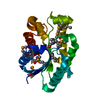
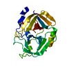
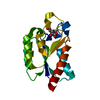
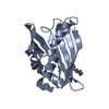
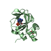
- PDB-5tv7: 2.05 Angstrom Resolution Crystal Structure of Peptidoglycan-Bindi... -
+ Open data

- Basic information
| Entry | Database: PDB / ID: 5tv7 | ||||||
|---|---|---|---|---|---|---|---|
| Title | 2.05 Angstrom Resolution Crystal Structure of Peptidoglycan-Binding Protein from Clostridioides difficile in Complex with Glutamine Hydroxamate. | ||||||
 Components Components | Putative peptidoglycan-binding/hydrolysing protein | ||||||
 Keywords Keywords | HYDROLASE / Structural Genomics / Center for Structural Genomics of Infectious Diseases / CSGID / Peptidoglycan-Binding Protein / Glutamine Hydroxamate | ||||||
| Function / homology | : / Putative peptidoglycan-binding domain / PGBD superfamily / Peptidoglycan binding-like / Putative peptidoglycan binding domain / PGBD-like superfamily / GLUTAMINE HYDROXAMATE / Peptidoglycan-binding/hydrolysing protein Function and homology information Function and homology information | ||||||
| Biological species |  Peptoclostridium difficile (bacteria) Peptoclostridium difficile (bacteria) | ||||||
| Method |  X-RAY DIFFRACTION / SYNCHROTRON / SAD / Resolution: 2.05 Å X-RAY DIFFRACTION / SYNCHROTRON / SAD / Resolution: 2.05 Å | ||||||
 Authors Authors | Minasov, G. / Wawrzak, Z. / Shuvalova, L. / Winsor, J. / Dubrovska, I. / Grimshaw, S. / Kwon, K. / Anderson, W.F. / Center for Structural Genomics of Infectious Diseases (CSGID) | ||||||
 Citation Citation | Journal: To Be Published Title: 2.05 Angstrom Resolution Crystal Structure of Peptidoglycan-Binding Protein from Clostridioides difficile in Complex with Glutamine Hydroxamate. Authors: Minasov, G. / Wawrzak, Z. / Shuvalova, L. / Winsor, J. / Dubrovska, I. / Grimshaw, S. / Kwon, K. / Anderson, W.F. / Center for Structural Genomics of Infectious Diseases (CSGID) | ||||||
| History |
|
- Structure visualization
| Structure viewer | Molecule: MolmilJmol/JSmol |
|---|
- Downloads & links
-Download
| PDBx/mmCIF format | 5tv7.cif.gz | 146.2 KB | Display | PDBx/mmCIF format |
|---|---|---|---|---|
| PDB format | pdb5tv7.ent.gz | 114.8 KB | Display | PDB format |
| PDBx/mmJSON format | 5tv7.json.gz | Tree view | PDBx/mmJSON format | |
| Others |  Other downloads Other downloads |
-Validation report
| Summary document | 5tv7_validation.pdf.gz | 453.7 KB | Display | wwPDB validaton report |
|---|---|---|---|---|
| Full document | 5tv7_full_validation.pdf.gz | 454.7 KB | Display | |
| Data in XML | 5tv7_validation.xml.gz | 16.4 KB | Display | |
| Data in CIF | 5tv7_validation.cif.gz | 22.7 KB | Display | |
| Arichive directory | https://data.pdbj.org/pub/pdb/validation_reports/tv/5tv7ftp://data.pdbj.org/pub/pdb/validation_reports/tv/5tv7 | HTTPS FTP |
-Related structure data
| Similar structure data | |
|---|---|
| Other databases |


-Links
 PDBj
PDBj
- Assembly
| Deposited unit | 
| ||||||||
|---|---|---|---|---|---|---|---|---|---|
| 1 | 
| ||||||||
| 2 | 
| ||||||||
| Unit cell |
|
-Components
| #1: Protein | Mass: 19749.502 Da / Num. of mol.: 2 Source method: isolated from a genetically manipulated source Source: (gene. exp.) Peptoclostridium difficile (strain 630) (bacteria)Strain: 630 / Gene: CD630_23880 / Plasmid: pMCSG7 / Production host: #2: Chemical | 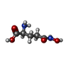  Mass: 162.144 Da / Num. of mol.: 2 / Source method: obtained synthetically / Formula: C5H10N2O4 Mass: 162.144 Da / Num. of mol.: 2 / Source method: obtained synthetically / Formula: C5H10N2O4#3: Water | ChemComp-HOH / |  Mass: 18.015 Da / Num. of mol.: 201 / Source method: isolated from a natural source / Formula: H2O Mass: 18.015 Da / Num. of mol.: 201 / Source method: isolated from a natural source / Formula: H2OHas protein modification | Y | |
|---|
-Experimental details
-Experiment
| Experiment | Method: X-RAY DIFFRACTION / Number of used crystals: 1 |
|---|
- Sample preparation
| Crystal | Density Matthews: 2.41 Å3/Da / Density % sol: 49.1 % |
|---|---|
| Crystal grow | Temperature: 295 K / Method: vapor diffusion, sitting drop / pH: 8.5 Details: Protein: 7.8 mg/ml, 0.25M Sodium chloride, 0.01M Tris HCl (pH 8.3), Screen: JCSG+ (B3), 0.1M Bicine (pH 8.5), 20% (w/v) PEG 6000. |
-Data collection
| Diffraction | Mean temperature: 100 K |
|---|---|
| Diffraction source | Source: SYNCHROTRON / Site: APS  / Beamline: 21-ID-F / Wavelength: 0.97872 Å / Beamline: 21-ID-F / Wavelength: 0.97872 Å |
| Detector | Type: MARMOSAIC 300 mm CCD / Detector: CCD / Date: Jun 4, 2014 / Details: C(111) |
| Radiation | Monochromator: beryllium lenses / Protocol: SINGLE WAVELENGTH / Monochromatic (M) / Laue (L): M / Scattering type: x-ray |
| Radiation wavelength | Wavelength: 0.97872 Å / Relative weight: 1 |
| Reflection | Resolution: 2.05→29.08 Å / Num. obs: 24487 / % possible obs: 100 % / Observed criterion σ(I): -3 / Redundancy: 5 % / Biso Wilson estimate: 32.2 Å2 / Rmerge(I) obs: 0.093 / Rsym value: 0.093 / Net I/σ(I): 28.1 |
| Reflection shell | Resolution: 2.05→2.09 Å / Redundancy: 5 % / Rmerge(I) obs: 0.769 / Mean I/σ(I) obs: 2.06 / CC1/2: 0.769 / % possible all: 99.9 |
- Processing
| Software |
| ||||||||||||||||||||||||||||||||||||||||||||||||||||||||||||||||||||||||||||||||||||||||||||||||||||||||||||||||||||||||||||||||||||||||||||||||||||||||||||||||||||||||||||||||||||||
|---|---|---|---|---|---|---|---|---|---|---|---|---|---|---|---|---|---|---|---|---|---|---|---|---|---|---|---|---|---|---|---|---|---|---|---|---|---|---|---|---|---|---|---|---|---|---|---|---|---|---|---|---|---|---|---|---|---|---|---|---|---|---|---|---|---|---|---|---|---|---|---|---|---|---|---|---|---|---|---|---|---|---|---|---|---|---|---|---|---|---|---|---|---|---|---|---|---|---|---|---|---|---|---|---|---|---|---|---|---|---|---|---|---|---|---|---|---|---|---|---|---|---|---|---|---|---|---|---|---|---|---|---|---|---|---|---|---|---|---|---|---|---|---|---|---|---|---|---|---|---|---|---|---|---|---|---|---|---|---|---|---|---|---|---|---|---|---|---|---|---|---|---|---|---|---|---|---|---|---|---|---|---|---|
| Refinement | Method to determine structure: SAD / Resolution: 2.05→29.08 Å / Cor.coef. Fo:Fc: 0.967 / Cor.coef. Fo:Fc free: 0.959 / SU B: 9.416 / SU ML: 0.123 / Cross valid method: THROUGHOUT / ESU R: 0.17 / ESU R Free: 0.151 / Details: HYDROGENS HAVE BEEN ADDED IN THE RIDING POSITIONS
| ||||||||||||||||||||||||||||||||||||||||||||||||||||||||||||||||||||||||||||||||||||||||||||||||||||||||||||||||||||||||||||||||||||||||||||||||||||||||||||||||||||||||||||||||||||||
| Solvent computation | Ion probe radii: 0.8 Å / Shrinkage radii: 0.8 Å / VDW probe radii: 1.2 Å | ||||||||||||||||||||||||||||||||||||||||||||||||||||||||||||||||||||||||||||||||||||||||||||||||||||||||||||||||||||||||||||||||||||||||||||||||||||||||||||||||||||||||||||||||||||||
| Displacement parameters | Biso mean: 48.198 Å2
| ||||||||||||||||||||||||||||||||||||||||||||||||||||||||||||||||||||||||||||||||||||||||||||||||||||||||||||||||||||||||||||||||||||||||||||||||||||||||||||||||||||||||||||||||||||||
| Refinement step | Cycle: 1 / Resolution: 2.05→29.08 Å
| ||||||||||||||||||||||||||||||||||||||||||||||||||||||||||||||||||||||||||||||||||||||||||||||||||||||||||||||||||||||||||||||||||||||||||||||||||||||||||||||||||||||||||||||||||||||
| Refine LS restraints |
|