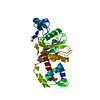
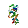
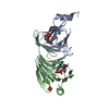
Entry Database : PDB / ID : 5tpvTitle X-ray structure of WlaRA (TDP-fucose-3,4-ketoisomerase) from Campylobacter jejuni WlaRA, TDP-fucose-3,4-ketoisomerase Keywords / / / / Function / homology Domain/homology Component
/ / / / / / / Biological species Campylobacter jejuni subsp. jejuni 81116 (Campylobacter)Method / / Resolution : 2.14 Å Authors Holden, H.M. / Thoden, J.B. / Li, Z.A. / Riegert, A.S. / Goneau, M.-F. / Cunningham, A.M. / Vinograd, E. / Schoenhofen, I.C. / Gilbert, M. / Li, J. Funding support Organization Grant number Country National Institutes of Health/National Institute of General Medical Sciences (NIH/NIGMS) GM115921
Journal : Glycobiology / Year : 2017Title : Characterization of the dTDP-Fuc3N and dTDP-Qui3N biosynthetic pathways in Campylobacter jejuni 81116.Authors : Li, Z.Z. / Riegert, A.S. / Goneau, M.F. / Cunningham, A.M. / Vinogradov, E. / Li, J. / Schoenhofen, I.C. / Thoden, J.B. / Holden, H.M. / Gilbert, M. History Deposition Oct 21, 2016 Deposition site / Processing site Revision 1.0 Feb 1, 2017 Provider / Type Revision 1.1 Apr 12, 2017 Group Revision 1.2 Sep 27, 2017 Group / Category / Item Revision 1.3 Apr 4, 2018 Group / Category / Item Revision 1.4 Apr 11, 2018 Group / Category / Item Revision 1.5 Jan 1, 2020 Group / Category / Item Revision 1.6 Oct 4, 2023 Group / Database references / Refinement descriptionCategory chem_comp_atom / chem_comp_bond ... chem_comp_atom / chem_comp_bond / database_2 / pdbx_initial_refinement_model Item / _database_2.pdbx_database_accession
Show all Show less
 Movie
Movie Controller

 Yorodumi
 Open data
 Basic information
 Components
 Keywords
 Function and homology information
 Campylobacter jejuni subsp. jejuni 81116 (Campylobacter)
 X-RAY DIFFRACTION /
  Authors
 United States, 1items
  Citation
 Structure visualization
 Downloads & links
 Other downloads
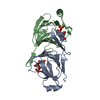


 PDBj
PDBj Assembly
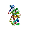


 Mass: 402.188 Da / Num. of mol.: 3 / Source method: obtained synthetically / Formula: C10H16N2O11P2
 Mass: 94.971 Da / Num. of mol.: 2 / Source method: obtained synthetically / Formula: PO4
 Mass: 18.015 Da / Num. of mol.: 211 / Source method: isolated from a natural source / Formula: H2O
 Sample preparation
 Processing