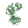
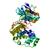
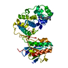
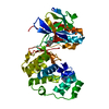
[English] 日本語
Yorodumi
- PDB-5tco: Human p38 MAP Kinase in Complex with Dibenzosuberone Compound 1 -

+
Open data


ID or keywords:

Loading...

-
Basic information

Entry
Database: PDB / ID: 5tco
TitleHuman p38 MAP Kinase in Complex with Dibenzosuberone Compound 1
ComponentsMitogen-activated protein kinase 14
KeywordsTRANSFERASE / Inhibitor / Dibenzosuberone / Human p38 MAP Kinase
Function / homology
Function and homology information


positive regulation of cyclase activity / stress-activated protein kinase signaling cascade / Activation of PPARGC1A (PGC-1alpha) by phosphorylation / CD163 mediating an anti-inflammatory response / regulation of synaptic membrane adhesion / stress-induced premature senescence / cell surface receptor protein serine/threonine kinase signaling pathway / 3'-UTR-mediated mRNA stabilization / KSRP (KHSRP) binds and destabilizes mRNA / cartilage condensation ...positive regulation of cyclase activity / stress-activated protein kinase signaling cascade / Activation of PPARGC1A (PGC-1alpha) by phosphorylation / CD163 mediating an anti-inflammatory response / regulation of synaptic membrane adhesion / stress-induced premature senescence / cell surface receptor protein serine/threonine kinase signaling pathway / 3'-UTR-mediated mRNA stabilization / KSRP (KHSRP) binds and destabilizes mRNA / cartilage condensation / DSCAM interactions / cellular response to UV-B / Platelet sensitization by LDL / mitogen-activated protein kinase p38 binding / positive regulation of myoblast fusion / positive regulation of muscle cell differentiation / negative regulation of hippo signaling / positive regulation of myotube differentiation / NFAT protein binding / Myogenesis / glucose import / Activation of the AP-1 family of transcription factors / regulation of cytokine production involved in inflammatory response / ERK/MAPK targets / p38MAPK cascade / fatty acid oxidation / cellular response to lipoteichoic acid / MAP kinase kinase activity / response to dietary excess / response to muramyl dipeptide / RHO GTPases Activate NADPH Oxidases / MAP kinase activity / regulation of ossification / cellular response to vascular endothelial growth factor stimulus / mitogen-activated protein kinase / signal transduction in response to DNA damage / positive regulation of myoblast differentiation / chondrocyte differentiation / vascular endothelial growth factor receptor signaling pathway / stress-activated MAPK cascade / skeletal muscle tissue development / positive regulation of cardiac muscle cell proliferation / lipopolysaccharide-mediated signaling pathway / negative regulation of inflammatory response to antigenic stimulus / p38MAPK events / striated muscle cell differentiation / response to muscle stretch / positive regulation of interleukin-12 production / positive regulation of brown fat cell differentiation / osteoclast differentiation / positive regulation of erythrocyte differentiation / DNA damage checkpoint signaling / activated TAK1 mediates p38 MAPK activation / cellular response to ionizing radiation / stem cell differentiation / positive regulation of glucose import / NOD1/2 Signaling Pathway / response to insulin / bone development / negative regulation of canonical Wnt signaling pathway / placenta development / cell morphogenesis / platelet activation / cellular response to virus / VEGFA-VEGFR2 Pathway / spindle pole / positive regulation of protein import into nucleus / osteoblast differentiation / ADP signalling through P2Y purinoceptor 1 / glucose metabolic process / chemotaxis / positive regulation of reactive oxygen species metabolic process / cellular senescence / cellular response to tumor necrosis factor / cellular response to lipopolysaccharide / peptidyl-serine phosphorylation / protein phosphatase binding / angiogenesis / secretory granule lumen / Oxidative Stress Induced Senescence / Regulation of TP53 Activity through Phosphorylation / ficolin-1-rich granule lumen / transcription by RNA polymerase II / cell surface receptor signaling pathway / intracellular signal transduction / nuclear speck / protein serine kinase activity / protein serine/threonine kinase activity / glutamatergic synapse / apoptotic process / Neutrophil degranulation / positive regulation of gene expression / regulation of transcription by RNA polymerase II / enzyme binding / signal transduction / positive regulation of transcription by RNA polymerase II / mitochondrion / extracellular region / nucleoplasm / ATP binding
Similarity search - Function
Mitogen-activated protein kinase 14 / Mitogen-activated protein (MAP) kinase p38-like / Mitogen-activated protein (MAP) kinase, conserved site / MAP kinase signature. / Transferase(Phosphotransferase) domain 1 / Transferase(Phosphotransferase); domain 1 / Phosphorylase Kinase; domain 1 / Phosphorylase Kinase; domain 1 / Protein kinase domain / Serine/Threonine protein kinases, catalytic domain ...Mitogen-activated protein kinase 14 / Mitogen-activated protein (MAP) kinase p38-like / Mitogen-activated protein (MAP) kinase, conserved site / MAP kinase signature. / Transferase(Phosphotransferase) domain 1 / Transferase(Phosphotransferase); domain 1 / Phosphorylase Kinase; domain 1 / Phosphorylase Kinase; domain 1 / Protein kinase domain / Serine/Threonine protein kinases, catalytic domain / Protein kinase, ATP binding site / Protein kinases ATP-binding region signature. / Protein kinase domain profile. / Protein kinase domain / Protein kinase-like domain superfamily / 2-Layer Sandwich / Orthogonal Bundle / Mainly Alpha / Alpha Beta
Similarity search - Domain/homology
Chem-79Q / Mitogen-activated protein kinase 14
Similarity search - Component
Biological speciesHomo sapiens (human)
MethodX-RAY DIFFRACTION / SYNCHROTRON / MOLECULAR REPLACEMENT / molecular replacement / Resolution: 2.1 Å
AuthorsMayer-Wrangowski, S.C. / Rauh, D.
CitationJournal: Angew. Chem. Int. Ed. Engl. / Year: 2017
Title: Optimized Target Residence Time: Type I1/2 Inhibitors for p38 alpha MAP Kinase with Improved Binding Kinetics through Direct Interaction with the R-Spine.
Authors: Wentsch, H.K. / Walter, N.M. / Buhrmann, M. / Mayer-Wrangowski, S. / Rauh, D. / Zaman, G.J.R. / Willemsen-Seegers, N. / Buijsman, R.C. / Henning, M. / Dauch, D. / Zender, L. / Laufer, S.
History
DepositionSep 15, 2016Deposition site: RCSB / Processing site: PDBE
Revision 1.0Apr 19, 2017Provider: repository / Type: Initial release
Revision 1.1May 3, 2017Group: Database references
Revision 1.2Jul 29, 2020Group: Data collection / Derived calculations / Structure summary
Category: chem_comp / entity ...chem_comp / entity / pdbx_chem_comp_identifier / pdbx_entity_nonpoly / struct_site / struct_site_gen
Item: _chem_comp.mon_nstd_flag / _chem_comp.name ..._chem_comp.mon_nstd_flag / _chem_comp.name / _chem_comp.type / _entity.pdbx_description / _pdbx_entity_nonpoly.name
Description: Carbohydrate remediation / Provider: repository / Type: Remediation
Revision 1.3May 8, 2024Group: Data collection / Database references / Structure summary
Category: chem_comp / chem_comp_atom ...chem_comp / chem_comp_atom / chem_comp_bond / database_2
Item: _chem_comp.pdbx_synonyms / _database_2.pdbx_DOI / _database_2.pdbx_database_accession

-
Structure visualization

Structure viewerMolecule:
MolmilJmol/JSmol

Downloads & links

-
Assembly

Deposited unit
A: Mitogen-activated protein kinase 14
hetero molecules


Theoretical massNumber of molelcules
Total (without water)42,4464
Polymers41,2691
Non-polymers1,1773
Water1,04558
1


  • Idetical with deposited unit
  • defined by software
TypeNameSymmetry operationNumber
identity operation1_555x,y,z1
Buried area1410 Å2
ΔGint13 kcal/mol
Surface area16080 Å2
MethodPISA
Unit cell
Length a, b, c (Å)66.070, 69.750, 74.730
Angle α, β, γ (deg.)90.000, 90.000, 90.000
Int Tables number19
Space group name H-MP212121

-
Components

#1: Protein Mitogen-activated protein kinase 14 / MAPK 14 / Cytokine suppressive anti-inflammatory drug-binding protein / CSBP / MAP kinase MXI2 / ...MAPK 14 / Cytokine suppressive anti-inflammatory drug-binding protein / CSBP / MAP kinase MXI2 / MAX-interacting protein 2 / Mitogen-activated protein kinase p38 alpha / MAP kinase p38 alpha / Stress-activated protein kinase 2a / SAPK2a


Mass: 41269.055 Da / Num. of mol.: 1
Source method: isolated from a genetically manipulated source
Source: (gene. exp.) Homo sapiens (human) / Gene: MAPK14, CSBP, CSBP1, CSBP2, CSPB1, MXI2, SAPK2A / Production host: Escherichia coli (E. coli)
References: UniProt: Q16539, mitogen-activated protein kinase
#2: Chemical ChemComp-79Q / 3-[(3-benzamido-4-fluoranyl-phenyl)amino]-~{N}-(2-morpholin-4-ylethyl)-11-oxidanylidene-5,6-dihydrodibenzo[1,2-~{d}:1',2'-~{f}][7]annulene-9-carboxamide


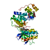
Mass: 592.659 Da / Num. of mol.: 1 / Source method: obtained synthetically / Formula: C35H33FN4O4
#3: Sugar ChemComp-BOG / octyl beta-D-glucopyranoside / Beta-Octylglucoside / octyl beta-D-glucoside / octyl D-glucoside / octyl glucoside


Type: D-saccharide / Mass: 292.369 Da / Num. of mol.: 2
Source method: isolated from a genetically manipulated source
Formula: C14H28O6 / Comment: detergent*YM
IdentifierTypeProgram
b-octylglucosideIUPAC CARBOHYDRATE SYMBOLPDB-CARE 1.0
#4: Water ChemComp-HOH / water


Mass: 18.015 Da / Num. of mol.: 58 / Source method: isolated from a natural source / Formula: H2O

-
Experimental details

-
Experiment

ExperimentMethod: X-RAY DIFFRACTION / Number of used crystals: 1

-
Sample preparation

CrystalDensity Matthews: 2.29 Å3/Da / Density % sol: 46.3 %
Crystal growTemperature: 293 K / Method: vapor diffusion, hanging drop / pH: 6.2 / Details: 100 mM MES, 22% PEG4000, 50 mM n-BOG

-
Data collection

DiffractionMean temperature: 90 K
Diffraction sourceSource: SYNCHROTRON / Site: SLS / Beamline: X10SA / Wavelength: 1 Å
DetectorType: PSI PILATUS 6M / Detector: PIXEL / Date: Sep 6, 2013
RadiationMonochromator: Si(111) / Protocol: SINGLE WAVELENGTH / Monochromatic (M) / Laue (L): M / Scattering type: x-ray
Radiation wavelengthWavelength: 1 Å / Relative weight: 1
ReflectionResolution: 2.1→50 Å / Num. obs: 20751 / % possible obs: 99.9 % / Redundancy: 6.51 % / Rrim(I) all: 0.077 / Net I/σ(I): 21.65
Reflection shellResolution: 2.1→2.2 Å / Mean I/σ(I) obs: 6.09 / Rrim(I) all: 0.402 / % possible all: 100

-
Phasing

PhasingMethod: molecular replacement

-
Processing

Software
NameVersionClassification
REFMAC5.7.0032refinement
XDSdata reduction
XSCALEdata scaling
PHASERphasing
RefinementMethod to determine structure: MOLECULAR REPLACEMENT / Resolution: 2.1→47.97 Å / Cor.coef. Fo:Fc: 0.933 / Cor.coef. Fo:Fc free: 0.895 / SU B: 6.064 / SU ML: 0.162 / Cross valid method: THROUGHOUT / σ(F): 0 / ESU R: 0.257 / ESU R Free: 0.215
RfactorNum. reflection% reflectionSelection details
Rfree0.2697 1038 5 %RANDOM
Rwork0.2145 ---
obs0.2172 19713 99.9 %-
Solvent computationIon probe radii: 0.8 Å / Shrinkage radii: 0.8 Å / VDW probe radii: 1.2 Å
Displacement parametersBiso max: 79.49 Å2 / Biso min: 13.07 Å2
Baniso -1Baniso -2Baniso -3
1-1.25 Å2-0 Å20 Å2
2--0.24 Å20 Å2
3----1.5 Å2
Refinement stepCycle: final / Resolution: 2.1→47.97 Å
ProteinNucleic acidLigandSolventTotal
Num. atoms2600 0 84 58 2742
Biso mean--33.36 26.47 -
Num. residues----327
Refine LS restraints
Refine-IDTypeDev idealDev ideal targetNumber
X-RAY DIFFRACTIONr_bond_refined_d0.0160.022747
X-RAY DIFFRACTIONr_bond_other_d0.0060.022627
X-RAY DIFFRACTIONr_angle_refined_deg1.8172.0013731
X-RAY DIFFRACTIONr_angle_other_deg0.9663.0095963
X-RAY DIFFRACTIONr_dihedral_angle_1_deg6.335323
X-RAY DIFFRACTIONr_dihedral_angle_2_deg36.6924.167120
X-RAY DIFFRACTIONr_dihedral_angle_3_deg14.215450
X-RAY DIFFRACTIONr_dihedral_angle_4_deg18.691515
X-RAY DIFFRACTIONr_chiral_restr0.0990.2425
X-RAY DIFFRACTIONr_gen_planes_refined0.0090.0213013
X-RAY DIFFRACTIONr_gen_planes_other0.0010.02618
X-RAY DIFFRACTIONr_mcbond_it2.272.6331304
X-RAY DIFFRACTIONr_mcbond_other2.2662.631303
X-RAY DIFFRACTIONr_mcangle_it3.3543.9291623
LS refinement shellResolution: 2.1→2.154 Å / Total num. of bins used: 20
RfactorNum. reflection% reflection
Rfree0.246 75 -
Rwork0.235 1420 -
all-1495 -
obs--99.93 %

+
About Yorodumi

-
News

-
Feb 9, 2022. New format data for meta-information of EMDB entries

New format data for meta-information of EMDB entries

  • Version 3 of the EMDB header file is now the official format.
  • The previous official version 1.9 will be removed from the archive.

Related info.:EMDB header

External links:wwPDB to switch to version 3 of the EMDB data model

-
Aug 12, 2020. Covid-19 info

Covid-19 info

URL: https://pdbjlvh1.pdbj.org/emnavi/covid19.php

New page: Covid-19 featured information page in EM Navigator.

Related info.:Covid-19 info / Mar 5, 2020. Novel coronavirus structure data

+
Mar 5, 2020. Novel coronavirus structure data

Novel coronavirus structure data

Related info.:Yorodumi Speices / Aug 12, 2020. Covid-19 info

External links:COVID-19 featured content - PDBj / Molecule of the Month (242):Coronavirus Proteases

+
Jan 31, 2019. EMDB accession codes are about to change! (news from PDBe EMDB page)

EMDB accession codes are about to change! (news from PDBe EMDB page)

  • The allocation of 4 digits for EMDB accession codes will soon come to an end. Whilst these codes will remain in use, new EMDB accession codes will include an additional digit and will expand incrementally as the available range of codes is exhausted. The current 4-digit format prefixed with “EMD-” (i.e. EMD-XXXX) will advance to a 5-digit format (i.e. EMD-XXXXX), and so on. It is currently estimated that the 4-digit codes will be depleted around Spring 2019, at which point the 5-digit format will come into force.
  • The EM Navigator/Yorodumi systems omit the EMD- prefix.

Related info.:Q: What is EMD? / ID/Accession-code notation in Yorodumi/EM Navigator

External links:EMDB Accession Codes are Changing Soon! / Contact to PDBj

+
Jul 12, 2017. Major update of PDB

Major update of PDB

  • wwPDB released updated PDB data conforming to the new PDBx/mmCIF dictionary.
  • This is a major update changing the version number from 4 to 5, and with Remediation, in which all the entries are updated.
  • In this update, many items about electron microscopy experimental information are reorganized (e.g. em_software).
  • Now, EM Navigator and Yorodumi are based on the updated data.

External links:wwPDB Remediation / Enriched Model Files Conforming to OneDep Data Standards Now Available in the PDB FTP Archive

-
Yorodumi

Thousand views of thousand structures

  • Yorodumi is a browser for structure data from EMDB, PDB, SASBDB, etc.
  • This page is also the successor to EM Navigator detail page, and also detail information page/front-end page for Omokage search.
  • The word "yorodu" (or yorozu) is an old Japanese word meaning "ten thousand". "mi" (miru) is to see.

Related info.:EMDB / PDB / SASBDB / Comparison of 3 databanks / Yorodumi Search / Aug 31, 2016. New EM Navigator & Yorodumi / Yorodumi Papers / Jmol/JSmol / Function and homology information / Changes in new EM Navigator and Yorodumi

Read more