butyryl-CoA catabolic process / propionyl-CoA metabolic process / propionyl-CoA catabolic process / coenzyme A diphosphatase / Peroxisomal lipid metabolism / malonyl-CoA catabolic process / medium-chain fatty-acyl-CoA catabolic process / coenzyme A diphosphatase activity / coenzyme A catabolic process / succinyl-CoA catabolic process ...butyryl-CoA catabolic process / propionyl-CoA metabolic process / propionyl-CoA catabolic process / coenzyme A diphosphatase / Peroxisomal lipid metabolism / malonyl-CoA catabolic process / medium-chain fatty-acyl-CoA catabolic process / coenzyme A diphosphatase activity / coenzyme A catabolic process / succinyl-CoA catabolic process / acetyl-CoA catabolic process / nucleoside diphosphate metabolic process / snoRNA binding / peroxisomal matrix / Hydrolases; Acting on acid anhydrides; In phosphorus-containing anhydrides / brown fat cell differentiation / Peroxisomal protein import / manganese ion binding / peroxisome / magnesium ion binding / cytosol Similarity search - Function
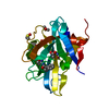
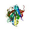
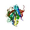
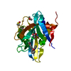
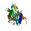
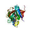
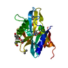
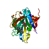
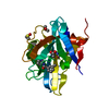
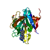
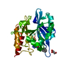
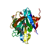
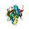
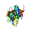
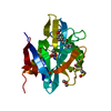
Mass: 22197.600 Da / Num. of mol.: 1 Source method: isolated from a genetically manipulated source Source: (gene. exp.) Homo sapiens (human) / Gene: NUDT7 / Production host: Escherichia coli (E. coli) References: UniProt: P0C024, Hydrolases; Acting on acid anhydrides; In phosphorus-containing anhydrides
 Movie
Movie Controller

 Yorodumi
 Open data
 Basic information
 Components
 Keywords
 Function and homology information
 Homo sapiens (human)
 X-RAY DIFFRACTION /
  Authors
 Citation
 Structure visualization
 Downloads & links
 Other downloads
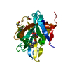


 PDBj
PDBj

 Assembly


 Mass: 59.044 Da / Num. of mol.: 2 / Source method: obtained synthetically / Formula: C2H3O2
 Mass: 261.316 Da / Num. of mol.: 1 / Source method: obtained synthetically / Formula: C15H19NO3
 Mass: 18.015 Da / Num. of mol.: 175 / Source method: isolated from a natural source / Formula: H2O
 Sample preparation
 / Beamline: I04-1 / Wavelength: 0.91587 Å
 Processing