All 364 structures of this series were generated by an automated refinement pipeline and, thus, not ...All 364 structures of this series were generated by an automated refinement pipeline and, thus, not refined to full convergence (e.g. since no manual (re-)building was performed, fragments are missing in the structural model even when present as indicated by the electron density)
Mass: 18.015 Da / Num. of mol.: 265 / Source method: isolated from a natural source / Formula: H2O
Has protein modification
Y
-
Experimental details
-
Experiment
Experiment
Method: X-RAY DIFFRACTION / Number of used crystals: 1
-
Sample preparation
Crystal
Density Matthews: 2.43 Å3/Da / Density % sol: 49.48 %
Crystal grow
Temperature: 290 K / Method: vapor diffusion, sitting drop / pH: 4.6 Details: 0.1 M ammonium acetate, 0.1 M sodium acetate, 24-30% PEG 4000; crystal obtained by streak-seeding and soaked with 90 mM of fragment 253 with the SMILES code C[C@@H](CN)N1C[C@H](C)O[C@H](C)C1
Resolution: 1.139→28.125 Å / SU ML: 0.07 / Cross valid method: THROUGHOUT / σ(F): 1.36 / Phase error: 12.72 / Stereochemistry target values: ML Details: All 364 structures of this series were generated by an automated refinement pipeline and, thus, not refined to full convergence (e.g. since no manual (re-)building was performed, fragments ...Details: All 364 structures of this series were generated by an automated refinement pipeline and, thus, not refined to full convergence (e.g. since no manual (re-)building was performed, fragments are missing in the structural model even when present as indicated by the electron density)
Rfactor
Num. reflection
% reflection
Selection details
Rfree
0.1448
5873
5 %
RANDOM
Rwork
0.1307
111587
-
-
obs
0.1314
117460
99.56 %
-
Solvent computation
Shrinkage radii: 0.9 Å / VDW probe radii: 1.11 Å / Solvent model: FLAT BULK SOLVENT MODEL
In the structure databanks used in Yorodumi, some data are registered as the other names, "COVID-19 virus" and "2019-nCoV". Here are the details of the virus and the list of structure data.
Jan 31, 2019. EMDB accession codes are about to change! (news from PDBe EMDB page)
EMDB accession codes are about to change! (news from PDBe EMDB page)
The allocation of 4 digits for EMDB accession codes will soon come to an end. Whilst these codes will remain in use, new EMDB accession codes will include an additional digit and will expand incrementally as the available range of codes is exhausted. The current 4-digit format prefixed with “EMD-” (i.e. EMD-XXXX) will advance to a 5-digit format (i.e. EMD-XXXXX), and so on. It is currently estimated that the 4-digit codes will be depleted around Spring 2019, at which point the 5-digit format will come into force.
The EM Navigator/Yorodumi systems omit the EMD- prefix.
Related info.:Q: What is EMD? / ID/Accession-code notation in Yorodumi/EM Navigator
Yorodumi is a browser for structure data from EMDB, PDB, SASBDB, etc.
This page is also the successor to EM Navigator detail page, and also detail information page/front-end page for Omokage search.
The word "yorodu" (or yorozu) is an old Japanese word meaning "ten thousand". "mi" (miru) is to see.
Related info.:EMDB / PDB / SASBDB / Comparison of 3 databanks / Yorodumi Search / Aug 31, 2016. New EM Navigator & Yorodumi / Yorodumi Papers / Jmol/JSmol / Function and homology information / Changes in new EM Navigator and Yorodumi
 Movie
Movie Controller

 Yorodumi
 Open data
 Basic information
 Components
 Keywords
 Function and homology information
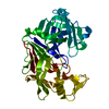
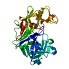
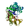
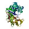
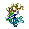
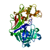
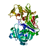
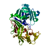
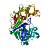
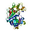
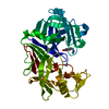
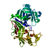
 Cryphonectria parasitica (chestnut blight fungus)
 X-RAY DIFFRACTION /
  Authors
 Citation
 Structure visualization
 Downloads & links
 Other downloads


 PDBj
PDBj
 Assembly
 Mass: 18.015 Da / Num. of mol.: 265 / Source method: isolated from a natural source / Formula: H2O
 Sample preparation
 / Beamline: 14.1 / Wavelength: 0.918409 Å
 Processing