[English] 日本語
Yorodumi
- PDB-5ow0: Crystal structure of an electron transfer flavoprotein from Geoba... -

+
Open data


ID or keywords:

Loading...

-
Basic information

Entry
Database: PDB / ID: 5ow0
Title
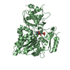
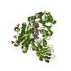
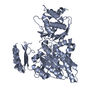
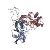
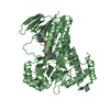
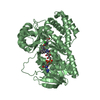
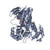
Crystal structure of an electron transfer flavoprotein from Geobacter metallireducens
Components
  • Electron transfer flavoprotein, alpha subunit
  • Electron transfer flavoprotein, beta subunit
KeywordsELECTRON TRANSPORT / anaerobic / toluene metabolism / FAD / AMP / flavoprotein
Function / homology
Function and homology information


flavin adenine dinucleotide binding / electron transfer activity
Similarity search - Function
Electron transfer flavoprotein, beta subunit / Electron transfer flavoprotein, beta subunit, N-terminal / Electron transfer flavoprotein alpha subunit/FixB / Electron transfer flavoprotein domain / Electron transfer flavoprotein, alpha/beta-subunit, N-terminal / Electron transfer flavoprotein, alpha subunit, C-terminal / Electron transfer flavoprotein FAD-binding domain / Electron transfer flavoprotein domain / TPP-binding domain / DHS-like NAD/FAD-binding domain superfamily ...Electron transfer flavoprotein, beta subunit / Electron transfer flavoprotein, beta subunit, N-terminal / Electron transfer flavoprotein alpha subunit/FixB / Electron transfer flavoprotein domain / Electron transfer flavoprotein, alpha/beta-subunit, N-terminal / Electron transfer flavoprotein, alpha subunit, C-terminal / Electron transfer flavoprotein FAD-binding domain / Electron transfer flavoprotein domain / TPP-binding domain / DHS-like NAD/FAD-binding domain superfamily / Rossmann-like alpha/beta/alpha sandwich fold / Rossmann fold / 3-Layer(aba) Sandwich / Alpha Beta
Similarity search - Domain/homology
ADENOSINE MONOPHOSPHATE / FLAVIN-ADENINE DINUCLEOTIDE / Electron transfer flavoprotein, alpha subunit / Electron transfer flavoprotein, beta subunit
Similarity search - Component
Biological speciesGeobacter metallireducens (bacteria)
MethodX-RAY DIFFRACTION / SYNCHROTRON / SAD / Resolution: 1.7 Å
AuthorsEssen, L.-O. / Vogt, M.S. / Heider, J. / Koelzer, S. / Peschke, P. / Chowdhury, N.P. / Schuehle, K. / Kleinsorge, D.
Funding support Germany, 1items
OrganizationGrant numberCountry
German Research FoundationSFB987/E09 Germany
CitationJournal: J.Bacteriol. / Year: 2019
Title: Structural and functional characterization of an electron transfer flavoprotein involved in toluene degradation in strictly anaerobic bacteria.
Authors: Vogt, M.S. / Schuhle, K. / Kolzer, S. / Peschke, P. / Chowdhury, N.P. / Kleinsorge, D. / Buckel, W. / Essen, L.O. / Heider, J.
History
DepositionAug 30, 2017Deposition site: PDBE / Processing site: PDBE
Revision 1.0Oct 10, 2018Provider: repository / Type: Initial release
Revision 1.1Sep 11, 2019Group: Data collection / Database references / Structure summary
Category: citation / citation_author ...citation / citation_author / entity / struct
Item: _citation.country / _citation.journal_abbrev ..._citation.country / _citation.journal_abbrev / _citation.journal_id_ASTM / _citation.journal_id_CSD / _citation.journal_id_ISSN / _citation.pdbx_database_id_DOI / _citation.pdbx_database_id_PubMed / _citation.title / _citation.year / _entity.formula_weight / _struct.title

-
Structure visualization

Structure viewerMolecule:
MolmilJmol/JSmol

Downloads & links

-
Assembly

Deposited unit
B: Electron transfer flavoprotein, beta subunit
A: Electron transfer flavoprotein, alpha subunit
hetero molecules


Theoretical massNumber of molelcules
Total (without water)59,6314
Polymers58,4982
Non-polymers1,1332
Water13,295738
1


  • Idetical with deposited unit
  • defined by software
  • Evidence: gel filtration
TypeNameSymmetry operationNumber
identity operation1_555x,y,z1
Buried area9120 Å2
ΔGint-57 kcal/mol
Surface area21640 Å2
MethodPISA
Unit cell
Length a, b, c (Å)54.730, 72.750, 133.881
Angle α, β, γ (deg.)90.00, 90.00, 90.00
Int Tables number19
Space group name H-MP212121

-
Components

#1: Protein Electron transfer flavoprotein, beta subunit


Mass: 27615.850 Da / Num. of mol.: 1
Source method: isolated from a genetically manipulated source
Source: (gene. exp.) Geobacter metallireducens (strain GS-15 / ATCC 53774 / DSM 7210) (bacteria)
Strain: GS-15 / ATCC 53774 / DSM 7210 / Gene: etfB-5, Gmet_1525 / Production host: Escherichia coli (E. coli) / References: UniProt: Q39VG5
#2: Protein Electron transfer flavoprotein, alpha subunit


Mass: 30881.887 Da / Num. of mol.: 1
Source method: isolated from a genetically manipulated source
Source: (gene. exp.) Geobacter metallireducens (strain GS-15 / ATCC 53774 / DSM 7210) (bacteria)
Strain: GS-15 / ATCC 53774 / DSM 7210 / Gene: etfA-5, Gmet_1526 / Production host: Escherichia coli (E. coli) / References: UniProt: Q39VG4
#3: Chemical ChemComp-AMP / ADENOSINE MONOPHOSPHATE


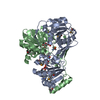
Mass: 347.221 Da / Num. of mol.: 1 / Source method: obtained synthetically / Formula: C10H14N5O7P / Comment: AMP*YM
#4: Chemical ChemComp-FAD / FLAVIN-ADENINE DINUCLEOTIDE


Mass: 785.550 Da / Num. of mol.: 1 / Source method: obtained synthetically / Formula: C27H33N9O15P2 / Comment: FAD*YM
#5: Water ChemComp-HOH / water


Mass: 18.015 Da / Num. of mol.: 738 / Source method: isolated from a natural source / Formula: H2O

-
Experimental details

-
Experiment

ExperimentMethod: X-RAY DIFFRACTION / Number of used crystals: 1

-
Sample preparation

CrystalDensity Matthews: 2.28 Å3/Da / Density % sol: 46.01 %
Crystal growTemperature: 277 K / Method: vapor diffusion, hanging drop
Details: Mother liquor: 15% (w/v) PEG 3350, 0.1 M HEPES, pH 7.5 mixed equal volumes of 10 mg/ml EtfAB in 300 mM NaCl, 20 mM HEPES, pH 7.5

-
Data collection

DiffractionMean temperature: 100 K
Diffraction sourceSource: SYNCHROTRON / Site: ESRF / Beamline: ID29 / Wavelength: 0.97929 Å
DetectorType: DECTRIS PILATUS3 6M / Detector: PIXEL / Date: Oct 8, 2015
RadiationProtocol: SINGLE WAVELENGTH / Monochromatic (M) / Laue (L): M / Scattering type: x-ray
Radiation wavelengthWavelength: 0.97929 Å / Relative weight: 1
ReflectionResolution: 1.7→44.63 Å / Num. obs: 59506 / % possible obs: 99.7 % / Redundancy: 12.8 % / Biso Wilson estimate: 17.44 Å2 / CC1/2: 0.998 / Rmerge(I) obs: 0.092 / Rpim(I) all: 0.038 / Net I/σ(I): 15.9
Reflection shellResolution: 1.7→1.73 Å / Redundancy: 2 % / Rmerge(I) obs: 0.584 / Mean I/σ(I) obs: 3.9 / Num. unique obs: 5846 / CC1/2: 0.918 / Rpim(I) all: 0.25 / % possible all: 99.6

-
Processing

Software
NameVersionClassification
PHENIX1.9_1692refinement
XDSdata reduction
Aimlessdata scaling
AutoSolphasing
RefinementMethod to determine structure: SAD / Resolution: 1.7→43.736 Å / SU ML: 0.14 / Cross valid method: FREE R-VALUE / σ(F): 1.34 / Phase error: 15.68
RfactorNum. reflection% reflection
Rfree0.1674 2970 5 %
Rwork0.1364 --
obs0.138 59434 99.66 %
Solvent computationShrinkage radii: 0.9 Å / VDW probe radii: 1.11 Å
Displacement parametersBiso mean: 24.1 Å2
Refinement stepCycle: LAST / Resolution: 1.7→43.736 Å
ProteinNucleic acidLigandSolventTotal
Num. atoms4053 0 76 738 4867
Refine LS restraints
Refine-IDTypeDev idealNumber
X-RAY DIFFRACTIONf_bond_d0.0154252
X-RAY DIFFRACTIONf_angle_d1.5395790
X-RAY DIFFRACTIONf_dihedral_angle_d14.5241606
X-RAY DIFFRACTIONf_chiral_restr0.066690
X-RAY DIFFRACTIONf_plane_restr0.008736
LS refinement shell
Resolution (Å)Rfactor RfreeNum. reflection RfreeRfactor RworkNum. reflection RworkRefine-ID% reflection obs (%)
1.7-1.72790.21341620.1962624X-RAY DIFFRACTION99
1.7279-1.75770.24831070.17632676X-RAY DIFFRACTION99
1.7577-1.78960.19891390.17082636X-RAY DIFFRACTION99
1.7896-1.82410.19121220.16662684X-RAY DIFFRACTION99
1.8241-1.86130.18971410.15692645X-RAY DIFFRACTION100
1.8613-1.90180.19411610.15512627X-RAY DIFFRACTION99
1.9018-1.9460.15281340.14492655X-RAY DIFFRACTION100
1.946-1.99470.18921280.14282645X-RAY DIFFRACTION99
1.9947-2.04860.18571550.142641X-RAY DIFFRACTION100
2.0486-2.10890.16561340.13472692X-RAY DIFFRACTION100
2.1089-2.1770.15351370.13542676X-RAY DIFFRACTION100
2.177-2.25480.16321270.13082682X-RAY DIFFRACTION100
2.2548-2.3450.17991570.12942668X-RAY DIFFRACTION100
2.345-2.45170.15311270.13232683X-RAY DIFFRACTION100
2.4517-2.5810.18581430.14052702X-RAY DIFFRACTION100
2.581-2.74270.19491640.13932689X-RAY DIFFRACTION100
2.7427-2.95440.19211250.1442712X-RAY DIFFRACTION100
2.9544-3.25160.16221390.13852735X-RAY DIFFRACTION100
3.2516-3.72190.15721530.12472718X-RAY DIFFRACTION100
3.7219-4.68840.12881330.11082784X-RAY DIFFRACTION100
4.6884-43.75010.15451820.13672883X-RAY DIFFRACTION100
Refinement TLS params.

Method: refined / Refine-ID: X-RAY DIFFRACTION

IDL112)L122)L132)L222)L232)L332)S11 (Å °)S12 (Å °)S13 (Å °)S21 (Å °)S22 (Å °)S23 (Å °)S31 (Å °)S32 (Å °)S33 (Å °)T112)T122)T132)T222)T232)T332)Origin x (Å)Origin y (Å)Origin z (Å)
11.3551-1.98590.04554.606-0.28421.12130.05660.10480.0206-0.3331-0.05130.0623-0.0414-0.11160.00160.125-0.0187-0.03050.1289-0.0060.084931.819930.996374.5179
24.6039-6.0522-3.12048.28943.61663.35630.1221-0.1540.2708-0.35360.0743-0.4339-0.09140.2333-0.17030.1704-0.01660.0310.1599-0.01740.175850.058424.616176.2034
31.04470.12880.02361.39640.15091.2547-0.02540.0224-0.0983-0.04740.016-0.02730.11020.05520.00650.12730.00340.01350.1089-0.00590.132640.460219.818281.4183
43.6094-0.46561.44688.46132.4244.1467-0.1261-0.11160.01550.26620.1435-0.325-0.00280.2134-0.02740.0966-0.00310.02060.18290.01020.09851.487926.09591.7234
51.48870.3046-1.6250.5397-0.76292.387-0.08980.3419-0.0361-0.30860.1232-0.09540.1228-0.0823-0.05080.16910.05710.00190.1657-0.00660.195544.57922.960967.9239
61.02981.4205-0.20194.6344-0.62260.4239-0.02610.03890.1265-0.1590.06070.1394-0.0244-0.061-0.03690.120.00810.00790.1524-0.00060.114232.206153.363181.9087
71.2548-0.56610.43520.9782-0.53251.9217-0.1387-0.1842-0.02960.18250.118-0.0005-0.0382-0.00350.02090.14790.00850.00760.14340.01260.123935.040530.5555103.8824
81.5873-2.55921.01135.1518-1.83310.6883-0.3411-0.06390.39560.71050.2312-1.0697-0.36640.1470.09370.2956-0.0206-0.07780.2627-0.05040.283353.455541.78795.4413
91.59880.1109-0.09832.4951-0.51051.16780.02290.00470.0735-0.04620.0261-0.1872-0.06360.0972-0.040.1072-0.01260.0060.0979-0.01870.091944.656853.279480.5803
Refinement TLS group
IDRefine-IDRefine TLS-IDSelection details
1X-RAY DIFFRACTION1chain 'B' and (resid 1 through 36 )
2X-RAY DIFFRACTION2chain 'B' and (resid 37 through 53 )
3X-RAY DIFFRACTION3chain 'B' and (resid 54 through 157 )
4X-RAY DIFFRACTION4chain 'B' and (resid 158 through 175 )
5X-RAY DIFFRACTION5chain 'B' and (resid 176 through 203 )
6X-RAY DIFFRACTION6chain 'B' and (resid 204 through 251 )
7X-RAY DIFFRACTION7chain 'A' and (resid 1 through 157 )
8X-RAY DIFFRACTION8chain 'A' and (resid 158 through 177 )
9X-RAY DIFFRACTION9chain 'A' and (resid 178 through 294 )

+
About Yorodumi

-
News

-
Feb 9, 2022. New format data for meta-information of EMDB entries

New format data for meta-information of EMDB entries

  • Version 3 of the EMDB header file is now the official format.
  • The previous official version 1.9 will be removed from the archive.

Related info.:EMDB header

External links:wwPDB to switch to version 3 of the EMDB data model

-
Aug 12, 2020. Covid-19 info

Covid-19 info

URL: https://pdbjlc1.pdbj.org/emnavi/covid19.php

New page: Covid-19 featured information page in EM Navigator.

Related info.:Covid-19 info / Mar 5, 2020. Novel coronavirus structure data

+
Mar 5, 2020. Novel coronavirus structure data

Novel coronavirus structure data

Related info.:Yorodumi Speices / Aug 12, 2020. Covid-19 info

External links:COVID-19 featured content - PDBj / Molecule of the Month (242):Coronavirus Proteases

+
Jan 31, 2019. EMDB accession codes are about to change! (news from PDBe EMDB page)

EMDB accession codes are about to change! (news from PDBe EMDB page)

  • The allocation of 4 digits for EMDB accession codes will soon come to an end. Whilst these codes will remain in use, new EMDB accession codes will include an additional digit and will expand incrementally as the available range of codes is exhausted. The current 4-digit format prefixed with “EMD-” (i.e. EMD-XXXX) will advance to a 5-digit format (i.e. EMD-XXXXX), and so on. It is currently estimated that the 4-digit codes will be depleted around Spring 2019, at which point the 5-digit format will come into force.
  • The EM Navigator/Yorodumi systems omit the EMD- prefix.

Related info.:Q: What is EMD? / ID/Accession-code notation in Yorodumi/EM Navigator

External links:EMDB Accession Codes are Changing Soon! / Contact to PDBj

+
Jul 12, 2017. Major update of PDB

Major update of PDB

  • wwPDB released updated PDB data conforming to the new PDBx/mmCIF dictionary.
  • This is a major update changing the version number from 4 to 5, and with Remediation, in which all the entries are updated.
  • In this update, many items about electron microscopy experimental information are reorganized (e.g. em_software).
  • Now, EM Navigator and Yorodumi are based on the updated data.

External links:wwPDB Remediation / Enriched Model Files Conforming to OneDep Data Standards Now Available in the PDB FTP Archive

-
Yorodumi

Thousand views of thousand structures

  • Yorodumi is a browser for structure data from EMDB, PDB, SASBDB, etc.
  • This page is also the successor to EM Navigator detail page, and also detail information page/front-end page for Omokage search.
  • The word "yorodu" (or yorozu) is an old Japanese word meaning "ten thousand". "mi" (miru) is to see.

Related info.:EMDB / PDB / SASBDB / Comparison of 3 databanks / Yorodumi Search / Aug 31, 2016. New EM Navigator & Yorodumi / Yorodumi Papers / Jmol/JSmol / Function and homology information / Changes in new EM Navigator and Yorodumi

Read more