 Movie
Movie Controller

[English] 日本語
 Yorodumi
- PDB-5oe8: Structure of large terminase from the thermophilic bacteriophage ... -
+ Open data

- Basic information
| Entry | Database: PDB / ID: 5oe8 | |||||||||
|---|---|---|---|---|---|---|---|---|---|---|
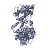
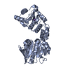
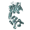
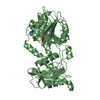
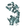
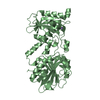
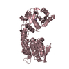
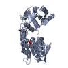
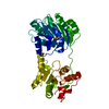
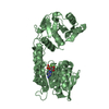
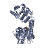
| Title | Structure of large terminase from the thermophilic bacteriophage D6E (Crystal form 2) | |||||||||
 Components Components | Large subunit terminase | |||||||||
 Keywords Keywords | VIRAL PROTEIN / large terminase | |||||||||
| Function / homology | Bacteriophage terminase, large subunit / Terminase large subunit, T4likevirus-type, N-terminal / nucleotide binding / P-loop containing nucleoside triphosphate hydrolase / metal ion binding / Large subunit terminase Function and homology information Function and homology information | |||||||||
| Biological species |  Deep-sea thermophilic phage D6E (virus) Deep-sea thermophilic phage D6E (virus) | |||||||||
| Method |  X-RAY DIFFRACTION / SYNCHROTRON / MOLECULAR REPLACEMENT / Resolution: 2.2 Å X-RAY DIFFRACTION / SYNCHROTRON / MOLECULAR REPLACEMENT / Resolution: 2.2 Å | |||||||||
 Authors Authors | Xu, R.G. / Jenkins, H.T. / Greive, S.J. / Antson, A.A. | |||||||||
| Funding support |  United Kingdom, 2items United Kingdom, 2items
| |||||||||
 Citation Citation | Journal: Nucleic Acids Res. / Year: 2017 Title: Structure of the large terminase from a hyperthermophilic virus reveals a unique mechanism for oligomerization and ATP hydrolysis. Authors: Xu, R.G. / Jenkins, H.T. / Antson, A.A. / Greive, S.J. | |||||||||
| History |
|
- Structure visualization
| Structure viewer | Molecule: MolmilJmol/JSmol |
|---|
- Downloads & links
-Download
| PDBx/mmCIF format | 5oe8.cif.gz | 269 KB | Display | PDBx/mmCIF format |
|---|---|---|---|---|
| PDB format | pdb5oe8.ent.gz | 215.2 KB | Display | PDB format |
| PDBx/mmJSON format | 5oe8.json.gz | Tree view | PDBx/mmJSON format | |
| Others |  Other downloads Other downloads |
-Validation report
| Arichive directory | https://data.pdbj.org/pub/pdb/validation_reports/oe/5oe8ftp://data.pdbj.org/pub/pdb/validation_reports/oe/5oe8 | HTTPS FTP |
|---|
-Related structure data


-Links
 PDBj
PDBj- Assembly
| Deposited unit | 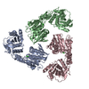
| ||||||||
|---|---|---|---|---|---|---|---|---|---|
| 1 | 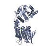
| ||||||||
| 2 | 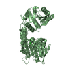
| ||||||||
| 3 | 
| ||||||||
| Unit cell |
| ||||||||
| Components on special symmetry positions |
|
-Components
| #1: Protein | Mass: 50166.492 Da / Num. of mol.: 3 Source method: isolated from a genetically manipulated source Source: (gene. exp.) Deep-sea thermophilic phage D6E (virus)Production host:  #2: Chemical | ChemComp-CL /   Mass: 35.453 Da / Num. of mol.: 6 / Source method: obtained synthetically / Formula: Cl Mass: 35.453 Da / Num. of mol.: 6 / Source method: obtained synthetically / Formula: Cl#3: Water | ChemComp-HOH / |  Mass: 18.015 Da / Num. of mol.: 769 / Source method: isolated from a natural source / Formula: H2O Mass: 18.015 Da / Num. of mol.: 769 / Source method: isolated from a natural source / Formula: H2O |
|---|
-Experimental details
-Experiment
| Experiment | Method: X-RAY DIFFRACTION / Number of used crystals: 1 |
|---|
- Sample preparation
| Crystal | Density Matthews: 2.84 Å3/Da / Density % sol: 56.66 % |
|---|---|
| Crystal grow | Temperature: 293 K / Method: vapor diffusion, sitting drop / Details: 0.1 M Ammonium Sulfate, 0.1 M HEPES pH 6.5-8.5. / PH range: 6.5-8.5 |
-Data collection
| Diffraction | Mean temperature: 100 K | ||||||||||||||||||||||||||||||
|---|---|---|---|---|---|---|---|---|---|---|---|---|---|---|---|---|---|---|---|---|---|---|---|---|---|---|---|---|---|---|---|
| Diffraction source | Source: SYNCHROTRON / Site: Diamond / Beamline: I04-1 / Wavelength: 0.9173 Å | ||||||||||||||||||||||||||||||
| Detector | Type: DECTRIS PILATUS 6M-F / Detector: PIXEL / Date: May 24, 2015 | ||||||||||||||||||||||||||||||
| Radiation | Protocol: SINGLE WAVELENGTH / Monochromatic (M) / Laue (L): M / Scattering type: x-ray | ||||||||||||||||||||||||||||||
| Radiation wavelength | Wavelength: 0.9173 Å / Relative weight: 1 | ||||||||||||||||||||||||||||||
| Reflection | Resolution: 2.2→46.05 Å / Num. obs: 80788 / % possible obs: 99.5 % / Redundancy: 4.2 % / CC1/2: 0.998 / Rmerge(I) obs: 0.08 / Rpim(I) all: 0.044 / Rrim(I) all: 0.091 / Net I/σ(I): 11.6 / Num. measured all: 335994 / Scaling rejects: 7 | ||||||||||||||||||||||||||||||
| Reflection shell | Diffraction-ID: 1
|
- Processing
| Software |
| ||||||||||||||||||||||||||||||||||||||||||||||||||||||||||||
|---|---|---|---|---|---|---|---|---|---|---|---|---|---|---|---|---|---|---|---|---|---|---|---|---|---|---|---|---|---|---|---|---|---|---|---|---|---|---|---|---|---|---|---|---|---|---|---|---|---|---|---|---|---|---|---|---|---|---|---|---|---|
| Refinement | Method to determine structure: MOLECULAR REPLACEMENT / Resolution: 2.2→46.05 Å / Cor.coef. Fo:Fc: 0.963 / Cor.coef. Fo:Fc free: 0.937 / SU B: 7.362 / SU ML: 0.173 / Cross valid method: THROUGHOUT / σ(F): 0 / ESU R: 0.23 / ESU R Free: 0.196 / Stereochemistry target values: MAXIMUM LIKELIHOOD Details: HYDROGENS HAVE BEEN ADDED IN THE RIDING POSITIONS U VALUES : REFINED INDIVIDUALLY
| ||||||||||||||||||||||||||||||||||||||||||||||||||||||||||||
| Solvent computation | Ion probe radii: 0.8 Å / Shrinkage radii: 0.8 Å / VDW probe radii: 1.2 Å / Solvent model: MASK | ||||||||||||||||||||||||||||||||||||||||||||||||||||||||||||
| Displacement parameters | Biso max: 114.98 Å2 / Biso mean: 50.295 Å2 / Biso min: 10 Å2
| ||||||||||||||||||||||||||||||||||||||||||||||||||||||||||||
| Refinement step | Cycle: final / Resolution: 2.2→46.05 Å
| ||||||||||||||||||||||||||||||||||||||||||||||||||||||||||||
| Refine LS restraints |
| ||||||||||||||||||||||||||||||||||||||||||||||||||||||||||||
| LS refinement shell | Resolution: 2.2→2.257 Å / Rfactor Rfree error: 0 / Total num. of bins used: 20
|
