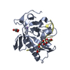
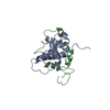
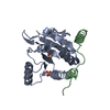
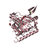
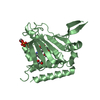
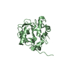
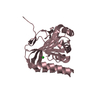
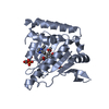
- PDB-5odj: Single-stranded DNA-binding protein from bacteriophage Enc34 -
+
Open data
ID or keywords:
Loading...
-
Basic information
Entry
Database: PDB / ID: 5odj
Title
Single-stranded DNA-binding protein from bacteriophage Enc34
Components
Single-stranded DNA-binding protein
Keywords
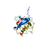
DNA BINDING PROTEIN / DNA replication / DUF2815 / SSB / OB fold
Function / homology
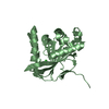
Enterobacter phage Enc34, ssDNA-binding protein / Enterobacter phage Enc34, ssDNA-binding protein / Nucleic acid-binding, OB-fold / metal ion binding / SsDNA-binding protein
Function and homology information
Biological species
Enterobacter phage Enc34 (virus)
Method
X-RAY DIFFRACTION / SYNCHROTRON / MAD / Resolution: 1.5 Å
 Movie
Movie Controller


 Open data
 Basic information
 Components
 Keywords
 Function and homology information
 Enterobacter phage Enc34 (virus)
 X-RAY DIFFRACTION /
  Authors
 Citation
 Structure visualization
 Downloads & links
 Other downloads
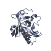
 PDBj
PDBj Assembly
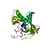


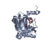
 Mass: 24.305 Da / Num. of mol.: 1 / Source method: obtained synthetically / Formula: Mg
 Mass: 35.453 Da / Num. of mol.: 1 / Source method: obtained synthetically / Formula: Cl
 Mass: 18.015 Da / Num. of mol.: 170 / Source method: isolated from a natural source / Formula: H2O
 Sample preparation
 / Beamline: I911-3 / Wavelength: 0.97671 Å
 Processing