 Movie
Movie Controller

[English] 日本語
 Yorodumi
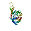
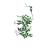
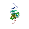
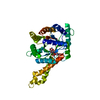
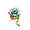
- PDB-5o9e: Crystal structure of the Imp4-Mpp10 complex from Chaetomium therm... -
+ Open data

- Basic information
| Entry | Database: PDB / ID: 5o9e | ||||||
|---|---|---|---|---|---|---|---|
| Title | Crystal structure of the Imp4-Mpp10 complex from Chaetomium thermophilum | ||||||
 Components Components |
| ||||||
 Keywords Keywords | RIBOSOME / Ribosome biogenesis / BRIX | ||||||
| Function / homology |  Function and homology information Function and homology informationMpp10 complex / rRNA primary transcript binding / sno(s)RNA-containing ribonucleoprotein complex / snoRNA binding / small-subunit processome / rRNA processing Similarity search - Function | ||||||
| Biological species |  Chaetomium thermophilum (fungus) Chaetomium thermophilum (fungus) | ||||||
| Method |  X-RAY DIFFRACTION / SYNCHROTRON / MOLECULAR REPLACEMENT / molecular replacement / Resolution: 1.884 Å X-RAY DIFFRACTION / SYNCHROTRON / MOLECULAR REPLACEMENT / molecular replacement / Resolution: 1.884 Å | ||||||
 Authors Authors | Kharde, S. / Ahmed, Y.L. / Sinning, I. | ||||||
 Citation Citation | Journal: PLoS ONE / Year: 2017 Title: Mpp10 represents a platform for the interaction of multiple factors within the 90S pre-ribosome. Authors: Sa-Moura, B. / Kornprobst, M. / Kharde, S. / Ahmed, Y.L. / Stier, G. / Kunze, R. / Sinning, I. / Hurt, E. | ||||||
| History |
|
- Structure visualization
| Structure viewer | Molecule: MolmilJmol/JSmol |
|---|
- Downloads & links
-Download
| PDBx/mmCIF format | 5o9e.cif.gz | 131.8 KB | Display | PDBx/mmCIF format |
|---|---|---|---|---|
| PDB format | pdb5o9e.ent.gz | 102.3 KB | Display | PDB format |
| PDBx/mmJSON format | 5o9e.json.gz | Tree view | PDBx/mmJSON format | |
| Others |  Other downloads Other downloads |
-Validation report
| Summary document | 5o9e_validation.pdf.gz | 441.4 KB | Display | wwPDB validaton report |
|---|---|---|---|---|
| Full document | 5o9e_full_validation.pdf.gz | 442.7 KB | Display | |
| Data in XML | 5o9e_validation.xml.gz | 13.7 KB | Display | |
| Data in CIF | 5o9e_validation.cif.gz | 18.8 KB | Display | |
| Arichive directory | https://data.pdbj.org/pub/pdb/validation_reports/o9/5o9eftp://data.pdbj.org/pub/pdb/validation_reports/o9/5o9e | HTTPS FTP |
-Related structure data
| Related structure data |  5by8S S: Starting model for refinement |
|---|---|
| Similar structure data |


-Links
 PDBj
PDBj
- Assembly
| Deposited unit | 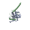
| ||||||||
|---|---|---|---|---|---|---|---|---|---|
| 1 |
| ||||||||
| Unit cell |
|
-Components
| #1: Protein | Mass: 22800.330 Da / Num. of mol.: 1 Source method: isolated from a genetically manipulated source Source: (gene. exp.) Chaetomium thermophilum (strain DSM 1495 / CBS 144.50 / IMI 039719) (fungus)Strain: DSM 1495 / CBS 144.50 / IMI 039719 / Gene: CTHT_0062900 / Plasmid: pET21g / Details (production host): modified pET21 / Production host:  |
|---|---|
| #2: Protein | Mass: 14787.353 Da / Num. of mol.: 1 Source method: isolated from a genetically manipulated source Source: (gene. exp.) Chaetomium thermophilum (strain DSM 1495 / CBS 144.50 / IMI 039719) (fungus)Strain: DSM 1495 / CBS 144.50 / IMI 039719 / Gene: CTHT_0046030 / Plasmid: pETZZ1a / Details (production host): modified pET24d / Production host: |
| #3: Chemical | ChemComp-EDO /   Mass: 62.068 Da / Num. of mol.: 1 / Source method: obtained synthetically / Formula: C2H6O2 Mass: 62.068 Da / Num. of mol.: 1 / Source method: obtained synthetically / Formula: C2H6O2 |
| #4: Water | ChemComp-HOH /  Mass: 18.015 Da / Num. of mol.: 136 / Source method: isolated from a natural source / Formula: H2O Mass: 18.015 Da / Num. of mol.: 136 / Source method: isolated from a natural source / Formula: H2O |
-Experimental details
-Experiment
| Experiment | Method: X-RAY DIFFRACTION / Number of used crystals: 1 |
|---|
- Sample preparation
| Crystal | Density Matthews: 2.15 Å3/Da / Density % sol: 42.88 % |
|---|---|
| Crystal grow | Temperature: 291 K / Method: vapor diffusion, sitting drop / Details: 20-30 % Ethylene glycol |
-Data collection
| Diffraction | Mean temperature: 100 K |
|---|---|
| Diffraction source | Source: SYNCHROTRON / Site: ESRF  / Beamline: ID23-2 / Wavelength: 0.8729 Å / Beamline: ID23-2 / Wavelength: 0.8729 Å |
| Detector | Type: DECTRIS PILATUS3 2M / Detector: PIXEL / Date: Jul 6, 2016 |
| Radiation | Protocol: SINGLE WAVELENGTH / Monochromatic (M) / Laue (L): M / Scattering type: x-ray |
| Radiation wavelength | Wavelength: 0.8729 Å / Relative weight: 1 |
| Reflection | Resolution: 1.88→48.883 Å / Num. obs: 26155 / % possible obs: 99.98 % / Redundancy: 7 % / Biso Wilson estimate: 27.64 Å2 / Net I/σ(I): 9.15 |
-Phasing
| Phasing | Method: molecular replacement |
|---|
- Processing
| Software |
| ||||||||||||||||||||||||||||||||||||||||||||||||||||||||||||||||||||||
|---|---|---|---|---|---|---|---|---|---|---|---|---|---|---|---|---|---|---|---|---|---|---|---|---|---|---|---|---|---|---|---|---|---|---|---|---|---|---|---|---|---|---|---|---|---|---|---|---|---|---|---|---|---|---|---|---|---|---|---|---|---|---|---|---|---|---|---|---|---|---|---|
| Refinement | Method to determine structure: MOLECULAR REPLACEMENT Starting model: 5BY8 Resolution: 1.884→48.883 Å / SU ML: 0.26 / Cross valid method: FREE R-VALUE / σ(F): 1.34 / Phase error: 22.68
| ||||||||||||||||||||||||||||||||||||||||||||||||||||||||||||||||||||||
| Solvent computation | Shrinkage radii: 0.9 Å / VDW probe radii: 1.11 Å | ||||||||||||||||||||||||||||||||||||||||||||||||||||||||||||||||||||||
| Displacement parameters | Biso max: 145.75 Å2 / Biso mean: 35.1536 Å2 / Biso min: 14.61 Å2 | ||||||||||||||||||||||||||||||||||||||||||||||||||||||||||||||||||||||
| Refinement step | Cycle: final / Resolution: 1.884→48.883 Å
| ||||||||||||||||||||||||||||||||||||||||||||||||||||||||||||||||||||||
| Refine LS restraints |
| ||||||||||||||||||||||||||||||||||||||||||||||||||||||||||||||||||||||
| LS refinement shell | Refine-ID: X-RAY DIFFRACTION / Rfactor Rfree error: 0 / Total num. of bins used: 9
| ||||||||||||||||||||||||||||||||||||||||||||||||||||||||||||||||||||||
| Refinement TLS params. | Method: refined / Origin x: 30.5593 Å / Origin y: 23.2614 Å / Origin z: 33.8023 Å
| ||||||||||||||||||||||||||||||||||||||||||||||||||||||||||||||||||||||
| Refinement TLS group |
|
