| Entry | Database: PDB / ID: 5o0x
|
|---|
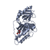
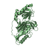
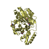
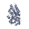
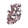
| Title | Crystal structure of dehydrogenase domain of Cylindrospermum stagnale NADPH-Oxidase 5 (NOX5) |
|---|
 Components Components | Putative ferric reductase |
|---|
 Keywords Keywords | OXIDOREDUCTASE / Membrane Protein / Reactive Oxygen Species / Oxidative Stress / Redox Biology |
|---|
| Function / homology |  Function and homology information Function and homology information
superoxide-generating NAD(P)H oxidase activity / NADPH oxidase complex / superoxide anion generation / defense response / nucleotide binding / calcium ion bindingSimilarity search - Function Ferric reductase, NAD binding domain / : / Ferric reductase NAD binding domain / FAD-binding 8 / FAD-binding domain / Ferric reductase transmembrane component-like domain / Ferric reductase like transmembrane component / Nucleotide-binding domain of ferredoxin-NADP reductase (FNR) module / EF-hand domain pair / FAD-binding domain, ferredoxin reductase-type ...Ferric reductase, NAD binding domain / : / Ferric reductase NAD binding domain / FAD-binding 8 / FAD-binding domain / Ferric reductase transmembrane component-like domain / Ferric reductase like transmembrane component / Nucleotide-binding domain of ferredoxin-NADP reductase (FNR) module / EF-hand domain pair / FAD-binding domain, ferredoxin reductase-type / Ferredoxin-NADP reductase (FNR), nucleotide-binding domain / Ferredoxin reductase-type FAD binding domain profile. / Riboflavin synthase-like beta-barrel / EF-hand domain pair / EF-hand, calcium binding motif / EF-Hand 1, calcium-binding site / EF-hand calcium-binding domain. / EF-hand calcium-binding domain profile. / EF-hand domain / EF-hand domain pair / Rossmann fold / 3-Layer(aba) Sandwich / Alpha BetaSimilarity search - Domain/homology |
|---|
| Biological species |  Cylindrospermum stagnale PCC 7417 (bacteria) Cylindrospermum stagnale PCC 7417 (bacteria) |
|---|
| Method |  X-RAY DIFFRACTION / SYNCHROTRON / MOLECULAR REPLACEMENT / Resolution: 2.2 Å X-RAY DIFFRACTION / SYNCHROTRON / MOLECULAR REPLACEMENT / Resolution: 2.2 Å |
|---|
 Authors Authors | Magnani, F. / Nenci, S. / Mattevi, A. |
|---|
| Funding support |  Italy, 1items Italy, 1items | Organization | Grant number | Country |
|---|
| Italian Ministry of Science and Education | PRIN2015-20152TE5PK_00 | Italy |
|
|---|
 Citation Citation | Journal: Proc. Natl. Acad. Sci. U.S.A. / Year: 2017
Title: Crystal structures and atomic model of NADPH oxidase.
Authors: Magnani, F. / Nenci, S. / Millana Fananas, E. / Ceccon, M. / Romero, E. / Fraaije, M.W. / Mattevi, A. |
|---|
| History | | Deposition | May 17, 2017 | Deposition site: PDBE / Processing site: PDBE |
|---|
| Revision 1.0 | Jun 28, 2017 | Provider: repository / Type: Initial release |
|---|
| Revision 1.1 | Jul 5, 2017 | Group: Database references / Category: citation
Item: _citation.country / _citation.journal_id_ASTM ..._citation.country / _citation.journal_id_ASTM / _citation.journal_id_CSD / _citation.journal_volume / _citation.page_first / _citation.page_last |
|---|
| Revision 1.2 | May 8, 2024 | Group: Data collection / Database references / Category: chem_comp_atom / chem_comp_bond / database_2
Item: _database_2.pdbx_DOI / _database_2.pdbx_database_accession |
|---|
|
|---|
 Movie
Movie Controller

 Yorodumi
 Open data
 Basic information
 Structure visualization
 Downloads & links
 Other downloads


 PDBj
PDBj

 Assembly
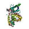


 Mass: 785.550 Da / Num. of mol.: 1 / Source method: obtained synthetically / Formula: C27H33N9O15P2 / Comment: FAD*YM
 Mass: 35.453 Da / Num. of mol.: 7 / Source method: obtained synthetically / Formula: Cl
 Mass: 282.331 Da / Num. of mol.: 5 / Source method: obtained synthetically / Formula: C12H26O7 / Comment: precipitant*YM
 Mass: 106.120 Da / Num. of mol.: 9 / Source method: obtained synthetically / Formula: C4H10O3
 Mass: 92.094 Da / Num. of mol.: 6 / Source method: obtained synthetically / Formula: C3H8O3
 Sample preparation
 / Beamline: ID29 / Wavelength: 0.8729 Å
 Processing