 Movie
Movie Controller

[English] 日本語
 Yorodumi
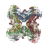
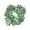
- PDB-5nb1: Crystal structures of homooligomers of collagen type IV. alpha4NC1 -
+ Open data

- Basic information
| Entry | Database: PDB / ID: 5nb1 | |||||||||
|---|---|---|---|---|---|---|---|---|---|---|
| Title | Crystal structures of homooligomers of collagen type IV. alpha4NC1 | |||||||||
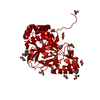
 Components Components | Collagen alpha-4(IV) chain | |||||||||
 Keywords Keywords | STRUCTURAL PROTEIN / Non-collagenous domain of collagen type IV. A principal structural component of basement membranes | |||||||||
| Function / homology |  Function and homology information Function and homology informationcollagen type IV trimer / Anchoring fibril formation / Crosslinking of collagen fibrils / glomerular basement membrane development / Collagen chain trimerization / extracellular matrix structural constituent conferring tensile strength / Collagen biosynthesis and modifying enzymes / Fibronectin matrix formation / Attachment of bacteria to epithelial cells / Signaling by PDGF ...collagen type IV trimer / Anchoring fibril formation / Crosslinking of collagen fibrils / glomerular basement membrane development / Collagen chain trimerization / extracellular matrix structural constituent conferring tensile strength / Collagen biosynthesis and modifying enzymes / Fibronectin matrix formation / Attachment of bacteria to epithelial cells / Signaling by PDGF / Laminin interactions / NCAM1 interactions / collagen fibril organization / Assembly of collagen fibrils and other multimeric structures / extracellular matrix structural constituent / Collagen degradation / Non-integrin membrane-ECM interactions / basement membrane / ECM proteoglycans / Integrin cell surface interactions / extracellular matrix / molecular adaptor activity / endoplasmic reticulum lumen / extracellular region Similarity search - Function | |||||||||
| Biological species |  Homo sapiens (human) Homo sapiens (human) | |||||||||
| Method |  X-RAY DIFFRACTION / SYNCHROTRON / MOLECULAR REPLACEMENT / molecular replacement / Resolution: 2.82 Å X-RAY DIFFRACTION / SYNCHROTRON / MOLECULAR REPLACEMENT / molecular replacement / Resolution: 2.82 Å | |||||||||
 Authors Authors | Casino, P. / Marina, A. | |||||||||
| Funding support |  Spain, 2items Spain, 2items
| |||||||||
 Citation Citation | Journal: IUCrJ / Year: 2018 Title: Structures of collagen IV globular domains: insight into associated pathologies, folding and network assembly. Authors: Casino, P. / Gozalbo-Rovira, R. / Rodriguez-Diaz, J. / Banerjee, S. / Boutaud, A. / Rubio, V. / Hudson, B.G. / Saus, J. / Cervera, J. / Marina, A. | |||||||||
| History |
|
- Structure visualization
| Structure viewer | Molecule: MolmilJmol/JSmol |
|---|
- Downloads & links
-Download
| PDBx/mmCIF format | 5nb1.cif.gz | 247.4 KB | Display | PDBx/mmCIF format |
|---|---|---|---|---|
| PDB format | pdb5nb1.ent.gz | 200.6 KB | Display | PDB format |
| PDBx/mmJSON format | 5nb1.json.gz | Tree view | PDBx/mmJSON format | |
| Others |  Other downloads Other downloads |
-Validation report
| Arichive directory | https://data.pdbj.org/pub/pdb/validation_reports/nb/5nb1ftp://data.pdbj.org/pub/pdb/validation_reports/nb/5nb1 | HTTPS FTP |
|---|
-Related structure data
| Related structure data |  5naxC  5nayC  5nazC  5nb0C 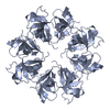 5nb2C 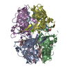 1t60S S: Starting model for refinement C: citing same article ( |
|---|---|
| Similar structure data |


-Links
 PDBj
PDBj


- Assembly
| Deposited unit | 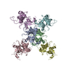
| ||||||||
|---|---|---|---|---|---|---|---|---|---|
| 1 | 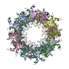
| ||||||||
| Unit cell |
|
-Components
| #1: Protein | Mass: 25443.912 Da / Num. of mol.: 6 Source method: isolated from a genetically manipulated source Source: (gene. exp.) Homo sapiens (human) / Gene: COL4A4 / Cell line (production host): Sf9 / Production host:   Spodoptera frugiperda (fall armyworm) / References: UniProt: P53420 Spodoptera frugiperda (fall armyworm) / References: UniProt: P53420#2: Water | ChemComp-HOH / |  Mass: 18.015 Da / Num. of mol.: 67 / Source method: isolated from a natural source / Formula: H2O Mass: 18.015 Da / Num. of mol.: 67 / Source method: isolated from a natural source / Formula: H2OHas protein modification | Y | |
|---|
-Experimental details
-Experiment
| Experiment | Method: X-RAY DIFFRACTION / Number of used crystals: 1 |
|---|
- Sample preparation
| Crystal | Density Matthews: 3.45 Å3/Da / Density % sol: 64.37 % |
|---|---|
| Crystal grow | Temperature: 294 K / Method: vapor diffusion, sitting drop Details: 6% PEG3350, 0.2 M sodium acetate and 0.1 M Mes pH 6.5 |
-Data collection
| Diffraction | Mean temperature: 100 K | ||||||||||||||||||||||||||||||||||||||||||||||||||||||||||||||||||||||||||||||||
|---|---|---|---|---|---|---|---|---|---|---|---|---|---|---|---|---|---|---|---|---|---|---|---|---|---|---|---|---|---|---|---|---|---|---|---|---|---|---|---|---|---|---|---|---|---|---|---|---|---|---|---|---|---|---|---|---|---|---|---|---|---|---|---|---|---|---|---|---|---|---|---|---|---|---|---|---|---|---|---|---|---|
| Diffraction source | Source: SYNCHROTRON / Site: ESRF  / Beamline: ID23-1 / Wavelength: 0.98 Å / Beamline: ID23-1 / Wavelength: 0.98 Å | ||||||||||||||||||||||||||||||||||||||||||||||||||||||||||||||||||||||||||||||||
| Detector | Type: RAYONIX MX-225 / Detector: CCD / Date: Jul 26, 2010 | ||||||||||||||||||||||||||||||||||||||||||||||||||||||||||||||||||||||||||||||||
| Radiation | Protocol: SINGLE WAVELENGTH / Monochromatic (M) / Laue (L): M / Scattering type: x-ray | ||||||||||||||||||||||||||||||||||||||||||||||||||||||||||||||||||||||||||||||||
| Radiation wavelength | Wavelength: 0.98 Å / Relative weight: 1 | ||||||||||||||||||||||||||||||||||||||||||||||||||||||||||||||||||||||||||||||||
| Reflection | Resolution: 2.82→47.464 Å / Num. all: 48121 / Num. obs: 48121 / % possible obs: 98.9 % / Redundancy: 5.4 % / Rpim(I) all: 0.04 / Rrim(I) all: 0.101 / Rsym value: 0.092 / Net I/av σ(I): 7.1 / Net I/σ(I): 12.7 / Num. measured all: 258124 | ||||||||||||||||||||||||||||||||||||||||||||||||||||||||||||||||||||||||||||||||
| Reflection shell | Diffraction-ID: 1
|
-Phasing
| Phasing | Method: molecular replacement |
|---|
- Processing
| Software |
| |||||||||||||||||||||||||||||||||||||||||||||
|---|---|---|---|---|---|---|---|---|---|---|---|---|---|---|---|---|---|---|---|---|---|---|---|---|---|---|---|---|---|---|---|---|---|---|---|---|---|---|---|---|---|---|---|---|---|---|
| Refinement | Method to determine structure: MOLECULAR REPLACEMENT Starting model: 1T60 Resolution: 2.82→47.46 Å / Cor.coef. Fo:Fc: 0.941 / Cor.coef. Fo:Fc free: 0.915 / SU B: 14.396 / SU ML: 0.266 / Cross valid method: THROUGHOUT / σ(F): 0 / ESU R: 0.181 / ESU R Free: 0.079 Details: HYDROGENS HAVE BEEN USED IF PRESENT IN THE INPUT U VALUES : REFINED INDIVIDUALLY
| |||||||||||||||||||||||||||||||||||||||||||||
| Solvent computation | Ion probe radii: 0.8 Å / Shrinkage radii: 0.8 Å / VDW probe radii: 1.2 Å | |||||||||||||||||||||||||||||||||||||||||||||
| Displacement parameters | Biso max: 147.78 Å2 / Biso mean: 56.903 Å2 / Biso min: 21.47 Å2
| |||||||||||||||||||||||||||||||||||||||||||||
| Refinement step | Cycle: final / Resolution: 2.82→47.46 Å
| |||||||||||||||||||||||||||||||||||||||||||||
| Refine LS restraints |
| |||||||||||||||||||||||||||||||||||||||||||||
| LS refinement shell | Resolution: 2.798→2.871 Å / Rfactor Rfree error: 0 / Total num. of bins used: 20
|
