 Movie
Movie Controller

[English] 日本語
 Yorodumi
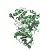
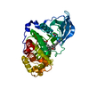
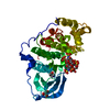
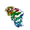
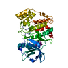
- PDB-5n9k: Crystal structure of human Protein kinase CK2 catalytic subunit i... -
+ Open data

- Basic information
| Entry | Database: PDB / ID: 5n9k | ||||||
|---|---|---|---|---|---|---|---|
| Title | Crystal structure of human Protein kinase CK2 catalytic subunit in complex with the ATP-competitive, tight-binding dibenzofuran inhibitor TF107 (5) | ||||||
 Components Components | Casein kinase II subunit alpha | ||||||
 Keywords Keywords | TRANSFERASE / Protein kinase / CK2 / Casein kinase 2 / Protein phosphorylation / ATP-competitive inhititors / dibenzofuran derivatives / TIGHT-BINDING INHIBITORS | ||||||
| Function / homology |  Function and homology information Function and homology informationPhosphorylation and nuclear translocation of BMAL1 (ARNTL) and CLOCK / positive regulation of aggrephagy / regulation of chromosome separation / WNT mediated activation of DVL / Condensation of Prometaphase Chromosomes / protein kinase CK2 complex / symbiont-mediated disruption of host cell PML body / Phosphorylation and nuclear translocation of the CRY:PER:kinase complex / Regulation of CDH1 posttranslational processing and trafficking to plasma membrane / Receptor Mediated Mitophagy ...Phosphorylation and nuclear translocation of BMAL1 (ARNTL) and CLOCK / positive regulation of aggrephagy / regulation of chromosome separation / WNT mediated activation of DVL / Condensation of Prometaphase Chromosomes / protein kinase CK2 complex / symbiont-mediated disruption of host cell PML body / Phosphorylation and nuclear translocation of the CRY:PER:kinase complex / Regulation of CDH1 posttranslational processing and trafficking to plasma membrane / Receptor Mediated Mitophagy / Synthesis of PC / Sin3-type complex / negative regulation of signal transduction by p53 class mediator / RUNX1 interacts with co-factors whose precise effect on RUNX1 targets is not known / Maturation of hRSV A proteins / negative regulation of apoptotic signaling pathway / negative regulation of double-strand break repair via homologous recombination / positive regulation of Wnt signaling pathway / negative regulation of proteasomal ubiquitin-dependent protein catabolic process / Signal transduction by L1 / Hsp90 protein binding / PML body / Wnt signaling pathway / Regulation of PTEN stability and activity / kinase activity / positive regulation of protein catabolic process / KEAP1-NFE2L2 pathway / rhythmic process / double-strand break repair / Cooperation of PDCL (PhLP1) and TRiC/CCT in G-protein beta folding / positive regulation of cell growth / protein folding / Regulation of TP53 Activity through Phosphorylation / non-specific serine/threonine protein kinase / regulation of cell cycle / negative regulation of translation / protein stabilization / protein serine kinase activity / protein serine/threonine kinase activity / apoptotic process / positive regulation of cell population proliferation / DNA damage response / signal transduction / nucleoplasm / ATP binding / identical protein binding / nucleus / plasma membrane / cytosol Similarity search - Function | ||||||
| Biological species |  Homo sapiens (human) Homo sapiens (human) | ||||||
| Method |  X-RAY DIFFRACTION / SYNCHROTRON / MOLECULAR REPLACEMENT / Resolution: 1.643 Å X-RAY DIFFRACTION / SYNCHROTRON / MOLECULAR REPLACEMENT / Resolution: 1.643 Å | ||||||
 Authors Authors | Schnitzler, A. / Gratz, A. / Bollacke, A. / Weyrich, M. / Kucklaender, U. / Wuensch, B. / Goetz, C. / Niefind, K. / Jose, J. | ||||||
 Citation Citation | Journal: Pharmaceuticals / Year: 2018 Title: A pi-Halogen Bond of Dibenzofuranones with the Gatekeeper Phe113 in Human Protein Kinase CK2 Leads to Potent Tight Binding Inhibitors. Authors: Schnitzler, A. / Gratz, A. / Bollacke, A. / Weyrich, M. / Kucklander, U. / Wunsch, B. / Gotz, C. / Niefind, K. / Jose, J. #1: Journal: J. Mol. Biol. / Year: 2003Title: Crystal structure of a C-terminal deletion mutant of human protein kinase CK2 catalytic subunit. Authors: Ermakova, I. / Boldyreff, B. / Issinger, O.G. / Niefind, K. #2: Journal: J. Mol. Biol. / Year: 2005Title: Inclining the purine base binding plane in protein kinase CK2 by exchanging the flanking side-chains generates a preference for ATP as a cosubstrate. Authors: Yde, C.W. / Ermakova, I. / Issinger, O.G. / Niefind, K. #3: Journal: Pharmaceuticals (Basel) / Year: 2017Title: Structural Hypervariability of the Two Human Protein Kinase CK2 Catalytic Subunit Paralogs Revealed by Complex Structures with a Flavonol- and a Thieno[2,3-d]pyrimidine-Based Inhibitor. Authors: Niefind, K. / Bischoff, N. / Golub, A.G. / Bdzhola, V.G. / Balanda, A.O. / Prykhod'ko, A.O. / Yarmoluk, S.M. #4: Journal: Biochim. Biophys. Acta / Year: 2012 Title: TF--a novel cell-permeable and selective inhibitor of human protein kinase CK2 induces apoptosis in the prostate cancer cell line LNCaP. Authors: Goetz, C. / Gratz, A. / Kucklaender, U. / Jose, J. | ||||||
| History |
|
- Structure visualization
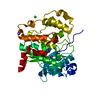
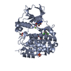
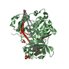
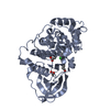
| Structure viewer | Molecule: MolmilJmol/JSmol |
|---|
- Downloads & links
-Download
| PDBx/mmCIF format | 5n9k.cif.gz | 163.6 KB | Display | PDBx/mmCIF format |
|---|---|---|---|---|
| PDB format | pdb5n9k.ent.gz | 128.8 KB | Display | PDB format |
| PDBx/mmJSON format | 5n9k.json.gz | Tree view | PDBx/mmJSON format | |
| Others |  Other downloads Other downloads |
-Validation report
| Arichive directory | https://data.pdbj.org/pub/pdb/validation_reports/n9/5n9kftp://data.pdbj.org/pub/pdb/validation_reports/n9/5n9k | HTTPS FTP |
|---|
-Related structure data
| Related structure data |  5n9lC  5n9nC  2pvrS C: citing same article ( S: Starting model for refinement |
|---|---|
| Similar structure data |


-Links
 PDBj
PDBj


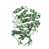
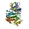
- Assembly
| Deposited unit | 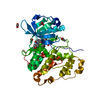
| ||||||||
|---|---|---|---|---|---|---|---|---|---|
| 1 |
| ||||||||
| Unit cell |
|
-Components
| #1: Protein | Mass: 40066.742 Da / Num. of mol.: 1 / Mutation: Deletion of C-terminal residues 336-391 Source method: isolated from a genetically manipulated source Source: (gene. exp.) Homo sapiens (human) / Gene: CSNK2A1, CK2A1 / Production host:  References: UniProt: P68400, non-specific serine/threonine protein kinase | ||||
|---|---|---|---|---|---|
| #2: Chemical | ChemComp-8QK /   Mass: 402.228 Da / Num. of mol.: 1 / Source method: obtained synthetically / Formula: C20H13Cl2NO4 Mass: 402.228 Da / Num. of mol.: 1 / Source method: obtained synthetically / Formula: C20H13Cl2NO4 | ||||
| #3: Chemical | ChemComp-ACT /   Mass: 59.044 Da / Num. of mol.: 6 / Source method: obtained synthetically / Formula: C2H3O2 Mass: 59.044 Da / Num. of mol.: 6 / Source method: obtained synthetically / Formula: C2H3O2#4: Chemical | ChemComp-GOL / |   Mass: 92.094 Da / Num. of mol.: 1 / Source method: obtained synthetically / Formula: C3H8O3 Mass: 92.094 Da / Num. of mol.: 1 / Source method: obtained synthetically / Formula: C3H8O3#5: Water | ChemComp-HOH / |  Mass: 18.015 Da / Num. of mol.: 249 / Source method: isolated from a natural source / Formula: H2O Mass: 18.015 Da / Num. of mol.: 249 / Source method: isolated from a natural source / Formula: H2O |
-Experimental details
-Experiment
| Experiment | Method: X-RAY DIFFRACTION / Number of used crystals: 1 |
|---|
- Sample preparation
| Crystal | Density Matthews: 1.96 Å3/Da / Density % sol: 37.29 % |
|---|---|
| Crystal grow | Temperature: 293 K / Method: vapor diffusion, sitting drop / pH: 5.6 Details: Prior to the crystallization the inhibitor was solubilized in 100 % DMSO in a concentration of 10 mM. Then, this inhibitor stock solution was mixed in a Ratio of 1:10 with human CK2alpha ...Details: Prior to the crystallization the inhibitor was solubilized in 100 % DMSO in a concentration of 10 mM. Then, this inhibitor stock solution was mixed in a Ratio of 1:10 with human CK2alpha (construct 1-335; solved with a Protein concentration of 8-10 mg/ml in 500 mM sodium chloride, 25 mM Tris/HCl pH 8.5). After a short time of incubation this mixture were mixed with reservoir solution [32 % (w/v) PEG4000, 0.2 M ammonium acetate, 0.1 M citrate pH 5.6] in a ratio of 2.5:1. 3.5 microliter of this final mixture was then equilibrated against the reservoir solution. The crystal growth was induced by seeding with 150 nanoliter seed suspension after an equilibration time of two days. |
-Data collection
| Diffraction | Mean temperature: 100 K |
|---|---|
| Diffraction source | Source: SYNCHROTRON / Site: SLS  / Beamline: X06DA / Wavelength: 0.99987 Å / Beamline: X06DA / Wavelength: 0.99987 Å |
| Detector | Type: DECTRIS PILATUS 2M / Detector: PIXEL / Date: Oct 12, 2012 |
| Radiation | Protocol: SINGLE WAVELENGTH / Monochromatic (M) / Laue (L): M / Scattering type: x-ray |
| Radiation wavelength | Wavelength: 0.99987 Å / Relative weight: 1 |
| Reflection | Resolution: 1.64→45.67 Å / Num. obs: 37660 / % possible obs: 99 % / Redundancy: 3.3 % / Rmerge(I) obs: 0.036 / Rsym value: 0.036 / Net I/σ(I): 17 |
| Reflection shell | Resolution: 1.64→1.67 Å / Redundancy: 2.8 % / Rmerge(I) obs: 0.645 / Mean I/σ(I) obs: 1.6 / Num. unique obs: 1606 / CC1/2: 0.716 / Rsym value: 0.645 / % possible all: 87 |
- Processing
| Software |
| |||||||||||||||||||||||||||||||||||||||||||||||||||||||||||||||||||||||||||||||||||||||||||||||||||||||||||||||||||||||||||||||||||||||||||||||||||||||||||||||||||||||||||||||
|---|---|---|---|---|---|---|---|---|---|---|---|---|---|---|---|---|---|---|---|---|---|---|---|---|---|---|---|---|---|---|---|---|---|---|---|---|---|---|---|---|---|---|---|---|---|---|---|---|---|---|---|---|---|---|---|---|---|---|---|---|---|---|---|---|---|---|---|---|---|---|---|---|---|---|---|---|---|---|---|---|---|---|---|---|---|---|---|---|---|---|---|---|---|---|---|---|---|---|---|---|---|---|---|---|---|---|---|---|---|---|---|---|---|---|---|---|---|---|---|---|---|---|---|---|---|---|---|---|---|---|---|---|---|---|---|---|---|---|---|---|---|---|---|---|---|---|---|---|---|---|---|---|---|---|---|---|---|---|---|---|---|---|---|---|---|---|---|---|---|---|---|---|---|---|---|---|
| Refinement | Method to determine structure: MOLECULAR REPLACEMENT Starting model: 2PVR Resolution: 1.643→36.22 Å / SU ML: 0.18 / Cross valid method: FREE R-VALUE / σ(F): 1.35 / Phase error: 19.76
| |||||||||||||||||||||||||||||||||||||||||||||||||||||||||||||||||||||||||||||||||||||||||||||||||||||||||||||||||||||||||||||||||||||||||||||||||||||||||||||||||||||||||||||||
| Solvent computation | Shrinkage radii: 0.9 Å / VDW probe radii: 1.11 Å | |||||||||||||||||||||||||||||||||||||||||||||||||||||||||||||||||||||||||||||||||||||||||||||||||||||||||||||||||||||||||||||||||||||||||||||||||||||||||||||||||||||||||||||||
| Refinement step | Cycle: LAST / Resolution: 1.643→36.22 Å
| |||||||||||||||||||||||||||||||||||||||||||||||||||||||||||||||||||||||||||||||||||||||||||||||||||||||||||||||||||||||||||||||||||||||||||||||||||||||||||||||||||||||||||||||
| Refine LS restraints |
| |||||||||||||||||||||||||||||||||||||||||||||||||||||||||||||||||||||||||||||||||||||||||||||||||||||||||||||||||||||||||||||||||||||||||||||||||||||||||||||||||||||||||||||||
| LS refinement shell |
| |||||||||||||||||||||||||||||||||||||||||||||||||||||||||||||||||||||||||||||||||||||||||||||||||||||||||||||||||||||||||||||||||||||||||||||||||||||||||||||||||||||||||||||||
| Refinement TLS params. | Method: refined / Refine-ID: X-RAY DIFFRACTION
| |||||||||||||||||||||||||||||||||||||||||||||||||||||||||||||||||||||||||||||||||||||||||||||||||||||||||||||||||||||||||||||||||||||||||||||||||||||||||||||||||||||||||||||||
| Refinement TLS group |
|
