 Movie
Movie Controller

[English] 日本語
 Yorodumi
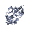
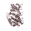
- PDB-5n7n: CRYSTAL STRUCTURE OF CATHEPSIN D ZYMOGEN FROM THE TICK IXODES RIC... -
+ Open data

- Basic information
| Entry | Database: PDB / ID: 5n7n | ||||||
|---|---|---|---|---|---|---|---|
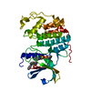
| Title | CRYSTAL STRUCTURE OF CATHEPSIN D ZYMOGEN FROM THE TICK IXODES RICINUS (IRCD1) | ||||||
 Components Components | Putative cathepsin d | ||||||
 Keywords Keywords | HYDROLASE | ||||||
| Function / homology |  Function and homology information Function and homology information | ||||||
| Biological species |  Ixodes ricinus (castor bean tick) Ixodes ricinus (castor bean tick) | ||||||
| Method |  X-RAY DIFFRACTION / SYNCHROTRON / FOURIER SYNTHESIS / Resolution: 2.3 Å X-RAY DIFFRACTION / SYNCHROTRON / FOURIER SYNTHESIS / Resolution: 2.3 Å | ||||||
 Authors Authors | Brynda, J. / Hanova, I. / Hobizalova, R. / Mares, M. | ||||||
 Citation Citation | Journal: Cell Chem Biol / Year: 2018 Title: Novel Structural Mechanism of Allosteric Regulation of Aspartic Peptidases via an Evolutionarily Conserved Exosite. Authors: Hanova, I. / Brynda, J. / Houstecka, R. / Alam, N. / Sojka, D. / Kopacek, P. / Maresova, L. / Vondrasek, J. / Horn, M. / Schueler-Furman, O. / Mares, M. | ||||||
| History |
|
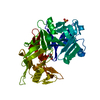
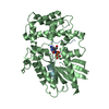
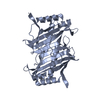
- Structure visualization
| Structure viewer | Molecule: MolmilJmol/JSmol |
|---|
- Downloads & links
-Download
| PDBx/mmCIF format | 5n7n.cif.gz | 282.7 KB | Display | PDBx/mmCIF format |
|---|---|---|---|---|
| PDB format | pdb5n7n.ent.gz | 229.3 KB | Display | PDB format |
| PDBx/mmJSON format | 5n7n.json.gz | Tree view | PDBx/mmJSON format | |
| Others |  Other downloads Other downloads |
-Validation report
| Arichive directory | https://data.pdbj.org/pub/pdb/validation_reports/n7/5n7nftp://data.pdbj.org/pub/pdb/validation_reports/n7/5n7n | HTTPS FTP |
|---|
-Related structure data


-Links
 PDBj
PDBj
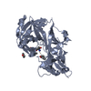
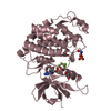
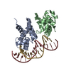
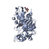
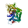
- Assembly
| Deposited unit | 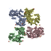
| ||||||||
|---|---|---|---|---|---|---|---|---|---|
| 1 | 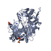
| ||||||||
| 2 | 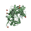
| ||||||||
| 3 | 
| ||||||||
| 4 | 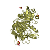
| ||||||||
| Unit cell |
|
-Components
| #1: Protein | Mass: 40225.188 Da / Num. of mol.: 4 Source method: isolated from a genetically manipulated source Source: (gene. exp.) Ixodes ricinus (castor bean tick) / Production host:  #2: Chemical | ChemComp-SO4 /   Mass: 96.063 Da / Num. of mol.: 18 / Source method: obtained synthetically / Formula: SO4 Mass: 96.063 Da / Num. of mol.: 18 / Source method: obtained synthetically / Formula: SO4#3: Chemical | ChemComp-NH4 /   Mass: 18.038 Da / Num. of mol.: 4 / Source method: obtained synthetically / Formula: H4N Mass: 18.038 Da / Num. of mol.: 4 / Source method: obtained synthetically / Formula: H4N#4: Water | ChemComp-HOH / |  Mass: 18.015 Da / Num. of mol.: 147 / Source method: isolated from a natural source / Formula: H2O Mass: 18.015 Da / Num. of mol.: 147 / Source method: isolated from a natural source / Formula: H2OHas protein modification | Y | |
|---|
-Experimental details
-Experiment
| Experiment | Method: X-RAY DIFFRACTION / Number of used crystals: 1 |
|---|
- Sample preparation
| Crystal | Density Matthews: 2.95 Å3/Da / Density % sol: 58.28 % |
|---|---|
| Crystal grow | Temperature: 291 K / Method: vapor diffusion, hanging drop / pH: 6.5 Details: 1:1 PROTEIN (10 MG/ML IN 2 MM BIS- TRIS, PH 6.5, 20 MM SODIUM CHLORIDE) TO RESERVOIR (0.085 M TRIS-HCL, PH 8.0, 1.7 M AMMONIUM SULFATE, 1 MM DTT), CRYOCOOLED IN MOTHER LIQUOR, VAPOR ...Details: 1:1 PROTEIN (10 MG/ML IN 2 MM BIS- TRIS, PH 6.5, 20 MM SODIUM CHLORIDE) TO RESERVOIR (0.085 M TRIS-HCL, PH 8.0, 1.7 M AMMONIUM SULFATE, 1 MM DTT), CRYOCOOLED IN MOTHER LIQUOR, VAPOR DIFFUSION, HANGING DROP, TEMPERATURE 291K |
-Data collection
| Diffraction | Mean temperature: 100 K |
|---|---|
| Diffraction source | Source: SYNCHROTRON / Site: ESRF  / Beamline: ID14-1 / Wavelength: 1.07161 Å / Beamline: ID14-1 / Wavelength: 1.07161 Å |
| Detector | Type: ADSC QUANTUM 315r / Detector: CCD / Date: Sep 30, 2013 |
| Radiation | Monochromator: CHANNEL CUT SI(111) / Protocol: SINGLE WAVELENGTH / Monochromatic (M) / Laue (L): M / Scattering type: x-ray |
| Radiation wavelength | Wavelength: 1.07161 Å / Relative weight: 1 |
| Reflection | Resolution: 2.3→45.87 Å / Num. obs: 82782 / % possible obs: 99.7 % / Observed criterion σ(I): -3 / Redundancy: 4.1 % / Rmerge(I) obs: 0.069 / Net I/σ(I): 14.81 |
| Reflection shell | Resolution: 2.3→2.44 Å / Rmerge(I) obs: 0.479 / Mean I/σ(I) obs: 2.79 / % possible all: 99.3 |
- Processing
| Software |
| ||||||||||||||||||||||||||||||||||||||||||||||||||||||||||||||||||||||||||||||||||||||||||||||||||||||||||||||||||||||||||||||||||||||||||||||||||||||||||||||||||||||||||||||||||||||
|---|---|---|---|---|---|---|---|---|---|---|---|---|---|---|---|---|---|---|---|---|---|---|---|---|---|---|---|---|---|---|---|---|---|---|---|---|---|---|---|---|---|---|---|---|---|---|---|---|---|---|---|---|---|---|---|---|---|---|---|---|---|---|---|---|---|---|---|---|---|---|---|---|---|---|---|---|---|---|---|---|---|---|---|---|---|---|---|---|---|---|---|---|---|---|---|---|---|---|---|---|---|---|---|---|---|---|---|---|---|---|---|---|---|---|---|---|---|---|---|---|---|---|---|---|---|---|---|---|---|---|---|---|---|---|---|---|---|---|---|---|---|---|---|---|---|---|---|---|---|---|---|---|---|---|---|---|---|---|---|---|---|---|---|---|---|---|---|---|---|---|---|---|---|---|---|---|---|---|---|---|---|---|---|
| Refinement | Method to determine structure: FOURIER SYNTHESIS / Resolution: 2.3→45.87 Å / Cor.coef. Fo:Fc: 0.936 / Cor.coef. Fo:Fc free: 0.906 / SU B: 7.849 / SU ML: 0.188 / Cross valid method: THROUGHOUT / ESU R: 0.297 / ESU R Free: 0.23 / Details: HYDROGENS HAVE BEEN ADDED IN THE RIDING POSITIONS
| ||||||||||||||||||||||||||||||||||||||||||||||||||||||||||||||||||||||||||||||||||||||||||||||||||||||||||||||||||||||||||||||||||||||||||||||||||||||||||||||||||||||||||||||||||||||
| Solvent computation | Ion probe radii: 0.8 Å / Shrinkage radii: 0.8 Å / VDW probe radii: 1.2 Å | ||||||||||||||||||||||||||||||||||||||||||||||||||||||||||||||||||||||||||||||||||||||||||||||||||||||||||||||||||||||||||||||||||||||||||||||||||||||||||||||||||||||||||||||||||||||
| Displacement parameters | Biso mean: 40.185 Å2
| ||||||||||||||||||||||||||||||||||||||||||||||||||||||||||||||||||||||||||||||||||||||||||||||||||||||||||||||||||||||||||||||||||||||||||||||||||||||||||||||||||||||||||||||||||||||
| Refinement step | Cycle: 1 / Resolution: 2.3→45.87 Å
| ||||||||||||||||||||||||||||||||||||||||||||||||||||||||||||||||||||||||||||||||||||||||||||||||||||||||||||||||||||||||||||||||||||||||||||||||||||||||||||||||||||||||||||||||||||||
| Refine LS restraints |
|