 Movie
Movie Controller

[English] 日本語
 Yorodumi
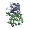
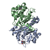
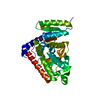
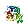
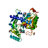
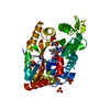
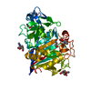
- PDB-5n53: Crystal structure of human 3-phosphoglycerate dehydrogenase in co... -
+ Open data

- Basic information
| Entry | Database: PDB / ID: 5n53 | ||||||
|---|---|---|---|---|---|---|---|
| Title | Crystal structure of human 3-phosphoglycerate dehydrogenase in complex with N-(3-chloro-4-methoxyphenyl) acetamide | ||||||
 Components Components | D-3-phosphoglycerate dehydrogenase | ||||||
 Keywords Keywords | OXIDOREDUCTASE / dehydrogenase / serine metabolism / FBDD | ||||||
| Function / homology |  Function and homology information Function and homology information2-oxoglutarate reductase / L-threonine metabolic process / glial cell development / taurine metabolic process / phosphoglycerate dehydrogenase / phosphoglycerate dehydrogenase activity / : / Serine metabolism / glycine metabolic process / (S)-malate dehydrogenase (NAD+, oxaloacetate-forming) ...2-oxoglutarate reductase / L-threonine metabolic process / glial cell development / taurine metabolic process / phosphoglycerate dehydrogenase / phosphoglycerate dehydrogenase activity / : / Serine metabolism / glycine metabolic process / (S)-malate dehydrogenase (NAD+, oxaloacetate-forming) / L-serine biosynthetic process / L-malate dehydrogenase (NAD+) activity / L-glutamine metabolic process / neural tube development / G1 to G0 transition / spinal cord development / brain development / NAD binding / neuron projection development / regulation of gene expression / electron transfer activity / extracellular exosome / cytosol Similarity search - Function | ||||||
| Biological species |  Homo sapiens (human) Homo sapiens (human) | ||||||
| Method |  X-RAY DIFFRACTION / SYNCHROTRON / MOLECULAR REPLACEMENT / Resolution: 1.48 Å X-RAY DIFFRACTION / SYNCHROTRON / MOLECULAR REPLACEMENT / Resolution: 1.48 Å | ||||||
 Authors Authors | Unterlass, J.E. / Basle, A. / Blackburn, T.J. / Tucker, J. / Cano, C. / Noble, M.E.M. / Curtin, N.J. | ||||||
| Funding support |  United Kingdom, 1items United Kingdom, 1items
| ||||||
 Citation Citation | Journal: Oncotarget / Year: 2018 Title: Validating and enabling phosphoglycerate dehydrogenase (PHGDH) as a target for fragment-based drug discovery in PHGDH-amplified breast cancer. Authors: Unterlass, J.E. / Basle, A. / Blackburn, T.J. / Tucker, J. / Cano, C. / Noble, M.E.M. / Curtin, N.J. | ||||||
| History |
|
- Structure visualization
| Structure viewer | Molecule: MolmilJmol/JSmol |
|---|
- Downloads & links
-Download
| PDBx/mmCIF format | 5n53.cif.gz | 176.7 KB | Display | PDBx/mmCIF format |
|---|---|---|---|---|
| PDB format | pdb5n53.ent.gz | 140.5 KB | Display | PDB format |
| PDBx/mmJSON format | 5n53.json.gz | Tree view | PDBx/mmJSON format | |
| Others |  Other downloads Other downloads |
-Validation report
| Arichive directory | https://data.pdbj.org/pub/pdb/validation_reports/n5/5n53ftp://data.pdbj.org/pub/pdb/validation_reports/n5/5n53 | HTTPS FTP |
|---|
-Related structure data
| Related structure data | 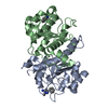 5nzoC  5nzpC 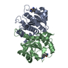 5nzqC 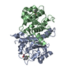 5ofvC 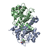 5ofwC 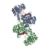 2g76S S: Starting model for refinement C: citing same article ( |
|---|---|
| Similar structure data |


-Links
 PDBj
PDBj
- Assembly
| Deposited unit | 
| ||||||||||||||||||
|---|---|---|---|---|---|---|---|---|---|---|---|---|---|---|---|---|---|---|---|
| 1 |
| ||||||||||||||||||
| Unit cell |
| ||||||||||||||||||
| Noncrystallographic symmetry (NCS) | NCS domain:
NCS domain segments: Component-ID: _ / Ens-ID: 1 / Beg auth comp-ID: ASN / Beg label comp-ID: ASN / End auth comp-ID: ARG / End label comp-ID: ARG / Refine code: _ / Auth seq-ID: 100 - 294 / Label seq-ID: 1 - 195
|
-Components
| #1: Protein | Mass: 20952.104 Da / Num. of mol.: 2 Source method: isolated from a genetically manipulated source Source: (gene. exp.) Homo sapiens (human) / Gene: PHGDH, PGDH3 / Production host:  #2: Chemical | 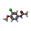  Mass: 199.634 Da / Num. of mol.: 2 / Source method: obtained synthetically / Formula: C9H10ClNO2 Mass: 199.634 Da / Num. of mol.: 2 / Source method: obtained synthetically / Formula: C9H10ClNO2#3: Water | ChemComp-HOH / |  Mass: 18.015 Da / Num. of mol.: 238 / Source method: isolated from a natural source / Formula: H2O Mass: 18.015 Da / Num. of mol.: 238 / Source method: isolated from a natural source / Formula: H2O |
|---|
-Experimental details
-Experiment
| Experiment | Method: X-RAY DIFFRACTION / Number of used crystals: 1 |
|---|
- Sample preparation
| Crystal | Density Matthews: 2.26 Å3/Da / Density % sol: 45.46 % |
|---|---|
| Crystal grow | Temperature: 298 K / Method: vapor diffusion, sitting drop / pH: 7 / Details: 0.1 M PCTP, pH 7, 23-25 % PEG 1500 |
-Data collection
| Diffraction | Mean temperature: 100 K |
|---|---|
| Diffraction source | Source: SYNCHROTRON / Site: Diamond / Beamline: I04-1 / Wavelength: 0.92 Å |
| Detector | Type: DECTRIS PILATUS3 S 6M / Detector: PIXEL / Date: Oct 22, 2014 |
| Radiation | Protocol: SINGLE WAVELENGTH / Monochromatic (M) / Laue (L): M / Scattering type: x-ray |
| Radiation wavelength | Wavelength: 0.92 Å / Relative weight: 1 |
| Reflection | Resolution: 1.48→50.12 Å / Num. obs: 51238 / % possible obs: 86.1 % / Redundancy: 2.13 % / Rmerge(I) obs: 0.111 / Rpim(I) all: 0.1 / Net I/σ(I): 4.6 |
| Reflection shell | Resolution: 1.5→1.53 Å / Redundancy: 2.14 % / Rmerge(I) obs: 1.088 / Mean I/σ(I) obs: 0.9 / Num. unique obs: 2530 / CC1/2: 0.355 / Rpim(I) all: 0.972 / % possible all: 85.8 |
- Processing
| Software |
| |||||||||||||||||||||||||||||||||||||||||||||||||||||||||||||||||||||||||||
|---|---|---|---|---|---|---|---|---|---|---|---|---|---|---|---|---|---|---|---|---|---|---|---|---|---|---|---|---|---|---|---|---|---|---|---|---|---|---|---|---|---|---|---|---|---|---|---|---|---|---|---|---|---|---|---|---|---|---|---|---|---|---|---|---|---|---|---|---|---|---|---|---|---|---|---|---|
| Refinement | Method to determine structure: MOLECULAR REPLACEMENT Starting model: 2G76 Resolution: 1.48→50.12 Å / Cor.coef. Fo:Fc: 0.954 / Cor.coef. Fo:Fc free: 0.925 / SU B: 5.83 / SU ML: 0.093 / Cross valid method: THROUGHOUT / σ(F): 0 / ESU R: 0.128 / ESU R Free: 0.102 Details: HYDROGENS HAVE BEEN ADDED IN THE RIDING POSITIONS U VALUES : REFINED INDIVIDUALLY
| |||||||||||||||||||||||||||||||||||||||||||||||||||||||||||||||||||||||||||
| Solvent computation | Ion probe radii: 0.8 Å / Shrinkage radii: 0.8 Å / VDW probe radii: 1.2 Å | |||||||||||||||||||||||||||||||||||||||||||||||||||||||||||||||||||||||||||
| Displacement parameters | Biso max: 101.98 Å2 / Biso mean: 19.681 Å2 / Biso min: 7.5 Å2
| |||||||||||||||||||||||||||||||||||||||||||||||||||||||||||||||||||||||||||
| Refinement step | Cycle: final / Resolution: 1.48→50.12 Å
| |||||||||||||||||||||||||||||||||||||||||||||||||||||||||||||||||||||||||||
| Refine LS restraints |
| |||||||||||||||||||||||||||||||||||||||||||||||||||||||||||||||||||||||||||
| Refine LS restraints NCS | Ens-ID: 1 / Number: 23386 / Refine-ID: X-RAY DIFFRACTION / Type: interatomic distance / Rms dev position: 0.07 Å / Weight position: 0.05
| |||||||||||||||||||||||||||||||||||||||||||||||||||||||||||||||||||||||||||
| LS refinement shell | Resolution: 1.484→1.522 Å / Rfactor Rfree error: 0 / Total num. of bins used: 20
|
