Mass: 18.015 Da / Num. of mol.: 319 / Source method: isolated from a natural source / Formula: H2O
-
Experimental details
-
Experiment
Experiment
Method: X-RAY DIFFRACTION / Number of used crystals: 1
-
Sample preparation
Crystal
Density Matthews: 2.1 Å3/Da / Density % sol: 42 %
Crystal grow
Temperature: 294 K / Method: vapor diffusion, sitting drop / pH: 9 Details: 1,54 M SODIUM CITRATE 60 MM TRIS- HCL, PH 9.0, PROTEIN 10 MG/ML 5-10 MM INHIBITOR (STOCK SOLUTION WAS 100 MM INHIBITOR DISSOLVED IN 100% DIMETHYL SULFOXIDE) VAPOR DIFFUSION, SITTING DROP, ...Details: 1,54 M SODIUM CITRATE 60 MM TRIS- HCL, PH 9.0, PROTEIN 10 MG/ML 5-10 MM INHIBITOR (STOCK SOLUTION WAS 100 MM INHIBITOR DISSOLVED IN 100% DIMETHYL SULFOXIDE) VAPOR DIFFUSION, SITTING DROP, TEMPERATURE 294K, TIME 2-5 DAYS
-
Data collection
Diffraction
Mean temperature: 100 K
Diffraction source
Source: SYNCHROTRON / Site: MAX II / Beamline: I911-3 / Wavelength: 0.97671 Å
 Movie
Movie Controller

 Yorodumi
 Open data
 Basic information
 Components
 Keywords
 Function and homology information
 Homo sapiens (human)
 X-RAY DIFFRACTION /
  Authors
 Citation
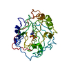
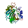
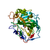
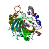
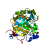
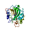
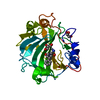
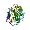
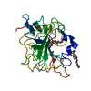
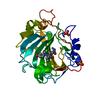
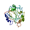
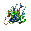
 Structure visualization
 Downloads & links
 Other downloads
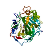


 PDBj
PDBj

 Assembly


 Mass: 65.409 Da / Num. of mol.: 1 / Source method: obtained synthetically / Formula: Zn
 Mass: 319.851 Da / Num. of mol.: 1 / Source method: obtained synthetically / Formula: C11H10ClNO2S3
 Mass: 18.015 Da / Num. of mol.: 319 / Source method: isolated from a natural source / Formula: H2O
 Sample preparation
 / Beamline: I911-3 / Wavelength: 0.97671 Å
 Processing